| Journal of Current Surgery, ISSN 1927-1298 print, 1927-1301 online, Open Access |
| Article copyright, the authors; Journal compilation copyright, J Curr Surg and Elmer Press Inc |
| Journal website https://jcs.elmerpub.com |
Original Article
Volume 16, Number 2, September 2026, pages 34-39

Serum Lactate Dehydrogenase as a Potential Biomarker for Lateral Lymph Node Metastasis in Thyroid Cancer Patients After Total Thyroidectomy
Fu Liang Suna, Zhi Qiang Suna, Jian Feng Chenga, Zhi Ming Songa, Qing Qing Huanga, b
aDepartment of Thyroid Surgery,Jiangsu Institute of Nuclear Medicine Jiangyuan Hospital of Jiangsu Province, Wuxi 214063, Jiangsu, China
bCorresponding Author: Qing Qing Huang, Jiangsu Institute of Nuclear Medicine Jiangyuan Hospital of Jiangsu Province, Wuxi 214063, Jiangsu, China
Manuscript submitted March 4, 2026, accepted June 30, 2026, published online August 29, 2026
Short title: LDH and Thyroid Lateral Lymph Node Metastasis
doi: https://doi.org/10.14740/jcs1035
| Abstract | ▴Top |
Background: This study aimed to investigate the clinical significance of serum lactate dehydrogenase (LDH) as a potential biomarker in patients with thyroid cancer, particularly in association with lateral lymph node metastasis (MLN) after total thyroidectomy.
Methods: A total of 2,560 patients with thyroid tumors were divided into benign group (n = 612) and malignant group (n = 1,948). Serum LDH and thyroid-stimulating hormone (TSH) levels were measured within 3 days before surgery. Within the malignant group, three subgroups were further analyzed: cancer subgroup, n = 1,820; MLN subgroup, n = 50; and cancer + MLN subgroup, n = 78. Statistical analyses included t-tests, Chi-square tests, and one-way analysis of variance (ANOVA) with Tukey’s post-hoc tests, multivariable logistic regression, and receiver operating characteristic (ROC) curve analysis.
Results: Compared with the benign group, patients in the malignant group were younger (t = 13.79, P < 0.001), had higher body mass index (BMI, t = 1.97, P = 0.049), a greater proportion of males (χ2 = 4.35, P = 0.037), and lower levels of both TSH (t = 6.72, P < 0.001) and LDH (t = 3.12, P = 0.002). Subgroup analysis within the malignant group revealed that the MLN subgroup had significantly higher LDH levels (197.9 ± 47.14 IU/L) compared with both the cancer subgroup (178.9 ± 45.58 IU/L, t = 2.90, P = 0.004) and the cancer + MLN subgroup (174.3 ± 46.49 IU/L, t = 2.79, P = 0.006). No significant difference was observed between the cancer and cancer + MLN subgroups (P = 0.58). Multivariable logistic regression confirmed that LDH remained independently associated with MLN status after adjusting for age, sex, and BMI (odds ratio (OR) = 1.011, 95% confidence interval (CI): 1.004–1.018, P = 0.002). ROC analysis yielded an area under the curve (AUC) of 0.628 (95% CI: 0.553–0.702), with an optimal cut-off of 187.5 IU/L (sensitivity 56.0%, specificity 63.6%).
Conclusions: While serum LDH levels are lower in patients with newly diagnosed thyroid cancer compared with those with benign tumors, a significant elevation in LDH is observed in patients who develop lateral cervical MLN after total thyroidectomy. These findings demonstrate an association between elevated serum LDH and lateral neck recurrence, suggesting that LDH may serve as a useful adjunctive biomarker for monitoring lateral MLN during postoperative follow-up in thyroid cancer patients. Further prospective studies with serial LDH measurements are warranted to validate these findings.
Keywords: Lactate dehydrogenase; Thyroid cancer; Postoperative surveillance; Lymph node metastasis
| Introduction | ▴Top |
Lactate dehydrogenase (LDH) is a key enzyme in the glycolytic pathway that catalyzes the interconversion of pyruvate and lactate. In tumor cells, even under aerobic conditions, glucose is preferentially metabolized via glycolysis—a phenomenon known as the Warburg effect—resulting in increased lactate production and LDH activity [1, 2]. This metabolic reprogramming supports rapid proliferation and creates an acidic microenvironment that promotes tumor progression and metastasis [3].
Serum LDH is an inexpensive and widely available laboratory parameter that has been investigated as a prognostic marker in various malignancies, including lymphoma, melanoma, and colorectal cancer [4–6]. In thyroid cancer, previous studies have demonstrated that LDHA, one of the LDH subunits, is overexpressed in papillary thyroid carcinoma (PTC) tissues and is associated with lymph node metastasis (MLN) and advanced stage [7]. However, clinical studies examining serum LDH levels in PTC patients remain limited and its potential role in postoperative surveillance—particularly for detecting lateral neck recurrence—has not been explored. Given that up to 37% of PTC patients may have lymph node metastases [8] and that lateral neck recurrence remains a clinical challenge, there is a need for accessible adjunctive biomarkers. Therefore, this study aimed to investigate the association between serum LDH levels in patients with PTC and lateral MLN after total thyroidectomy.
| Materials and Methods | ▴Top |
Study population
Patients with thyroid tumors who underwent surgery at our hospital from March 1, 2019, to May 1, 2021, were consecutively enrolled. Inclusion criteria were: (1) confirmed diagnosis by paraffin-embedded pathological examination; (2) serum LDH measurement performed within 3 days before surgery. Exclusion criteria included: abnormal liver or kidney function; history of myocardial infarction or hemolytic diseases within 3 months; active hepatitis; coexisting other malignancies; acute inflammation; poorly controlled blood glucose (fasting blood glucose > 10 mmol/L); and hyperthyroidism with abnormal thyroid function tests (FT3/FT4 above normal range) despite medication.
The enrolled patients were divided into two groups based on pathological diagnosis: a benign group (n = 612) and a malignant group (n = 1,948). The malignant group was further subdivided into three subgroups: (1) Cancer subgroup (n = 1,820): Patients with pathologically confirmed PTC, with or without central MLN, who underwent initial thyroid surgery. (2) MLN subgroup (n = 50): Patients who had previously undergone total thyroidectomy for PTC and were subsequently diagnosed with lateral cervical MLN during follow-up. Diagnosis of lateral MLN was confirmed by fine-needle aspiration cytology and/or surgical pathology. For these patients, serum LDH was measured at the time of metastasis diagnosis (before any intervention for metastasis). (3) Cancer + MLN subgroup (n = 78): Patients initially diagnosed with PTC accompanied by lateral cervical MLN at the time of their first surgery.
This study was performed in line with the principles of the Declaration of Helsinki. This study was approved by the Ethics Committee of Jiangsu Institute of Nuclear Medicine (No. YLK202515).
Laboratory measurements
Fasting peripheral venous blood samples were collected from all enrolled patients within 3 days before surgery. Serum LDH levels (reference range: 90–180 IU/L) were measured using an automatic biochemical analyzer with a colorimetric method. Thyroid-stimulating hormone (TSH) levels (reference range: 0.27–4.2 mIU/L) were measured using electrochemiluminescence immunoassay (Roche Diagnostics, Switzerland). Demographic information including age, sex, height, and weight was also collected, and body mass index (BMI) was calculated as weight (kg)/height (m2).
Statistical analysis
This study was a retrospective analysis incorporating biomedical data. Data distribution was assessed using the Shapiro–Wilk test; all continuous variables approximated normal distribution in this large sample, supporting the use of parametric tests. Homogeneity of variances was tested using Levene’s test.
Quantitative data are expressed as mean ± standard deviation (x ± s). Statistical analysis was performed using GraphPad Prism 8 software. The independent samples t-test was used to compare differences in LDH levels, TSH levels, age, and BMI between the benign and malignant groups, while the Chi-square test was used to analyze differences in gender composition. For comparisons involving multiple subgroups within the malignant group, one-way analysis of variance (ANOVA) was applied, followed by post-hoc comparisons using Tukey’s honest significant difference (HSD) test. To evaluate the independent association of LDH with MLN status, multivariable binary logistic regression was performed with MLN status (1 = MLN, 0 = cancer) as the dependent variable, and LDH, age, sex, and BMI as covariates. Receiver operating characteristic (ROC) curve analysis was conducted to evaluate the diagnostic performance of LDH in discriminating between MLN and cancer subgroups. The optimal cut-off value was determined using Youden’s index. TSH data were missing in 35 benign and 172 malignant cases due to incomplete laboratory orders; these cases were excluded from TSH analysis. The significance level was set at α = 0.05, and a P-value < 0.05 was considered statistically significant.
| Results | ▴Top |
Baseline characteristics of the two groups
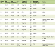
A total of 2,560 patients were included in the analysis, comprising 612 in the benign group and 1,948 in the malignant group. As shown in Table 1, patients in the malignant group were significantly younger, had a higher BMI, and included a greater proportion of males compared with those in the benign group (all P < 0.05).
 Click to view | Table 1. Baseline Characteristics of the Study Population (x ± s) |
TSH and LDH levels in benign and malignant groups
As shown in Table 2, both TSH and LDH levels were significantly lower in the malignant group compared with the benign group (P < 0.001 and P = 0.002, respectively). Notably, the mean LDH level in the benign group (185.90 ± 47.16 IU/L) was slightly above the upper limit of the normal reference range (180 IU/L), while the mean level in the malignant group (179.20 ± 45.74 IU/L) fell within the normal range. TSH data were available for 577 benign cases (94.3%) and 1,776 malignant cases (91.2%).
Click to view | Table 2. Comparison of TSH and LDH Levels Between Groups (x ± s) |
Subgroup analysis of LDH levels in malignant group
Subgroup analysis within the malignant group revealed significant differences in LDH levels among the three subgroups (one-way ANOVA: F = 5.67, P = 0.004). As shown in Figure 1, the MLN subgroup had significantly higher LDH levels (197.9 ± 47.14 IU/L) compared with both the cancer subgroup (178.9 ± 45.58 IU/L, t = 2.90, P = 0.004) and the cancer + MLN subgroup (174.3 ± 46.49 IU/L, t = 2.79, P = 0.006). No significant difference was observed between the cancer and cancer + MLN subgroups (P = 0.58).
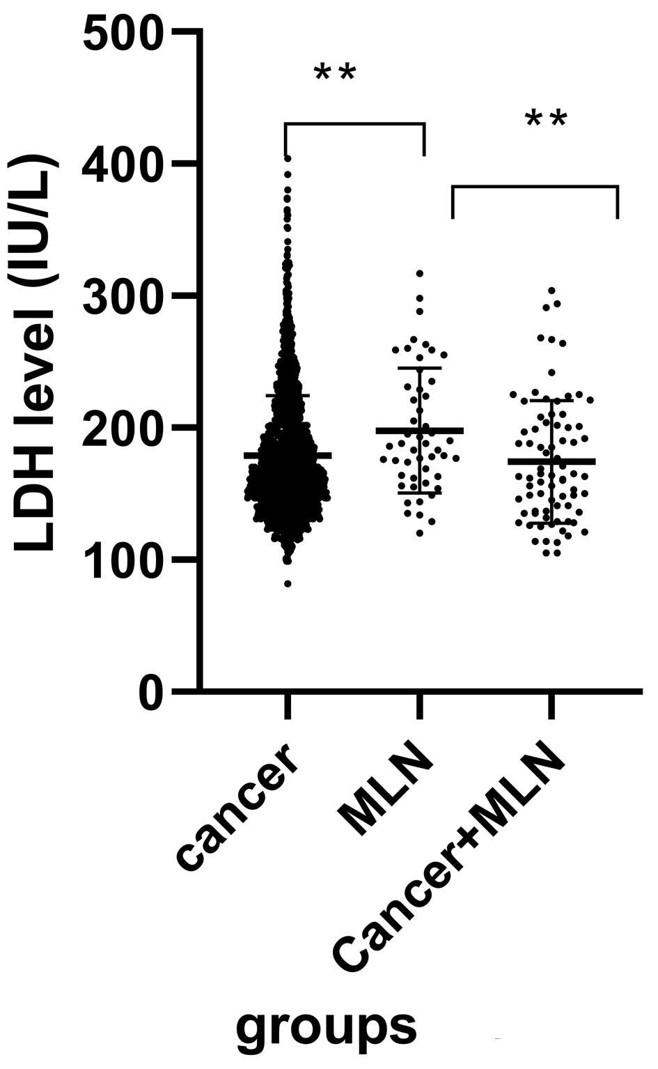 Click for large image | Figure 1. Serum lactate dehydrogenase (LDH) levels in subgroups of patients with malignant thyroid tumors (**P < 0.01). |
Multivariable logistic regression and ROC curve analysis
In the ROC analysis comparing the MLN subgroup (n = 50) with the cancer subgroup (n = 1,820), the cancer + MLN subgroup was excluded. Serum LDH yielded an area under the curve (AUC) of 0.628 (95% confidence interval (CI): 0.553–0.702, P = 0.002) (Fig. 2). The optimal cut-off value based on Youden’s index was 187.5 IU/L, with a sensitivity of 56.0% and a specificity of 63.6%.
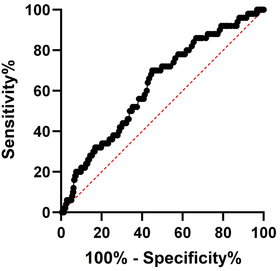 Click for large image | Figure 2. Receiver operating characteristic (ROC) curve analysis of serum lactate dehydrogenase (LDH) levels for distinguishing the MLN subgroup from the cancer subgroup. |
After adjusting for age, sex, and BMI, LDH remained independently associated with MLN status (odds ratio (OR) = 1.011, 95% CI: 1.004–1.018, P = 0.002) (Table 3). This indicates that for every 1 IU/L increase in serum LDH, the odds of being in the MLN subgroup increase by approximately 1.1%, independent of age, sex, and BMI.
Click to view | Table 3. Multivariable Logistic Regression Analysis for Factors Associated With MLN Status |
| Discussion | ▴Top |
Thyroid cancer is one of the tumors with the most rapidly increasing incidence worldwide, with PTC accounting for approximately 90% of cases. The 2015 American Thyroid Association (ATA) guidelines classify patients with ≤ 5 central lymph node micrometastases (< 2 mm) as low-risk in recurrence stratification. However, data show that up to 37% of PTC patients may present with lymph node metastases [8]. A multicenter study from China on intermediate- to high-risk differentiated thyroid cancer indicated that pathological examination after central neck dissection revealed MLN in as high as 82.3% [9]. Therefore, more sensitive and accessible biomarkers are needed to assist in preoperative planning and postoperative surveillance.
LDH, a key enzyme in glycolysis, has been implicated in tumor progression across multiple cancer types. Previous studies have demonstrated that LDHA, one of the subunits of LDH, is significantly overexpressed at both transcriptional and translational levels in PTC compared with normal thyroid tissue, and can induce epithelial–mesenchymal transition and autophagy, promoting PTC proliferation and metastasis [10]. Ban et al also confirmed high expression of LDHA in PTC, which was associated with MLN and advanced disease stage [11]. Additionally, Wang et al reported that HYOU1 can up-regulate LDHB mRNA expression in PTC cell lines, promoting glycolysis and tumor growth [12]. Given these findings, serum LDH has emerged as a potential diagnostic and prognostic marker for various malignancies, including thyroid cancer [13]. However, clinical studies specifically examining serum LDH levels in PTC patients remain limited.
In the present study, we found that patients in the malignant group had significantly lower serum LDH levels than those in the benign group, with the mean value in the benign group slightly exceeding the normal reference range. This finding differs from reports in more aggressive malignancies such as colorectal and lung cancer, where elevated LDH correlates with tumor burden and poor prognosis [14]. Several factors may explain this discrepancy. First, PTC is generally indolent and slowly progressive, which may result in lower glycolytic activity and less LDH release into the circulation compared with rapidly proliferating tumors. Second, the benign group included patients with subclinical hypothyroidism or hypothyroidism, conditions known to elevate serum LDH [15], potentially contributing to the higher LDH levels observed in this group. Third, the malignant group was significantly younger and had higher BMI, though the influence of age and BMI on LDH remains unclear. Regarding TSH, we observed significantly lower levels in the malignant group. While elevated TSH is a risk factor for thyroid nodule malignancy, this finding likely reflects the inclusion of patients on TSH-suppressive therapy. Importantly, the lower TSH in the malignant group does not explain the lower LDH, as hypothyroidism (which elevates LDH) was more common in the benign group. Thus, the LDH differences between groups appear to be driven more by tumor biology than by thyroid function status. Although statistically significant, the absolute difference in LDH between benign and malignant groups was modest (6.7 IU/L) and both means were within or near the normal reference range, limiting its diagnostic utility in individual patients.
The most important finding of this study emerges from the subgroup analysis within the malignant group. Patients who developed lateral cervical MLN after total thyroidectomy (the MLN subgroup) had significantly elevated serum LDH levels compared with both patients with cancer alone and patients initially presenting with lateral metastasis. Notably, this elevation was specific to the post-thyroidectomy recurrence setting—patients with initial lateral metastasis at diagnosis (cancer + MLN subgroup) did not show elevated LDH. This dissociation suggests two possible mechanisms: (1) Metabolic reprogramming during recurrence: Emerging evidence indicates that metastatic lesions can undergo metabolic adaptation, often exhibiting enhanced glycolytic activity compared with primary tumors [16, 17]. In PTC, recurrent disease in the lateral neck may be more glycolytically active, leading to increased LDH release. (2) Tumor burden effect: Even if not massive, the cumulative burden of recurrent disease might trigger a systemic metabolic response detectable by serum LDH. Distinguishing between these mechanisms requires further investigation, but both support the clinical utility of LDH as a monitoring tool.
Multivariable analysis confirmed LDH as an independent factor associated with MLN status. Although its diagnostic accuracy is only moderate (see Results), LDH remains clinically useful as a low-cost, widely available trigger biomarker. A rising LDH level during follow-up may prompt intensified surveillance with lateral neck ultrasound, even before imaging detects structural recurrence.
The clinical implications are practical and actionable. Serum LDH is routinely measured in standard laboratory panels at minimal cost. For thyroid cancer patients who have undergone total thyroidectomy, a rising LDH level—particularly if it exceeds the normal range or shows a consistent upward trend—may warrant enhanced surveillance, such as high-resolution ultrasound of the lateral neck compartments or more frequent follow-up visits. Conversely, a stable or declining LDH level may provide reassurance. However, LDH should not replace established tools (ultrasound, thyroglobulin) but rather serve as an adjunctive trigger for earlier or more targeted investigation.
This study has several limitations. First, as a retrospective single-center study, it is subject to inherent selection bias. Second, we did not measure LDH isoenzymes or tissue expression of LDHA/LDHB, which might provide additional mechanistic insights. Third, the sample size of the MLN subgroup (n = 50), while sufficient for statistical analysis, is relatively small, and these findings should be validated in larger, multicenter cohorts. Fourth, we did not collect data on dynamic changes in LDH over time within individual patients due to clinical practice. Without longitudinal measurements, it is not possible to determine whether LDH rose before recurrence, rose concurrently, or was unrelated to recurrence. Fifth, the MLN subgroup differed from other subgroups in terms of disease duration and prior treatment history, which may confound LDH comparisons. The MLN subgroup may represent biologically more aggressive tumors with inherently enhanced glycolytic activity, which could explain elevated LDH rather than the recurrence event itself. Additionally, due to the retrospective nature and the reliance on early handwritten surgical records, we were unable to include critical pathological variables such as tumor size, stage, extrathyroidal extension, histologic subtype, and vascular invasion in our multivariable regression model. These factors are important determinants of tumor aggressiveness. Prospective studies with serial LDH measurements are needed to establish the predictive value of LDH trajectories—specifically, to determine the sensitivity and specificity of a rising LDH for detecting lateral neck recurrence.
Conclusion
In summary, this study demonstrates that while serum LDH levels in patients with newly diagnosed thyroid cancer are lower than those in patients with benign thyroid tumors, a significant elevation in LDH is observed in patients who develop lateral cervical MLN after total thyroidectomy. Multivariable analysis confirms that this association is independent of age, sex, and BMI. However, the moderate diagnostic performance of LDH suggests that its value lies primarily as a low-cost adjunctive monitoring tool rather than a replacement for established surveillance methods. Serum LDH may therefore serve as a useful, cost-effective adjunctive biomarker for monitoring lateral MLN during postoperative follow-up in thyroid cancer patients. Further prospective studies with serial LDH measurements and comprehensive pathological data are warranted to validate these findings and to explore the underlying mechanisms linking LDH elevation to metastatic progression.
Acknowledgments
None to declare.
Financial Disclosure
The authors declare that no funds, grants, or other support were received during the preparation of this manuscript.
Conflict of Interest
The authors declare that they have no conflict of interest.
Informed Consent
As the research involved a retrospective analysis of de-identified data, the requirement for informed consent was waived.
Author Contributions
Zhi Qiang Sun contributed to conceptualization; Jian Feng Cheng to methodology; Zhi Ming Song to formal analysis and investigation; Fu Liang Sun to writing – original draft preparation; Qing Qing Huang to writing – review and editing. All authors read and approved the final manuscript.
Data Availability
The data that support the findings of this study are available from the corresponding author upon reasonable request.
| References | ▴Top |
- Vaupel P, Multhoff G. Revisiting the Warburg effect: historical dogma versus current understanding. J Physiol. 2021;599(6):1745-1757.
doi pubmed - Jurisic V, Radenkovic S, Konjevic G. The actual role of LDH as tumor marker, biochemical and clinical aspects. Adv Exp Med Biol. 2015;867:115-124.
doi pubmed - Papaneophytou C. The Warburg effect: is it always an enemy? Front Biosci (Landmark Ed). 2024;29(12):402.
doi pubmed - Hirami Y, Nishimura MF, Urata T, Morimoto M, Maekawa Y, Yoshino T, Nishimura Y, et al. Comparison of serum sIL-2R and LDH levels in patients with intravascular large B-cell lymphoma and patients with advanced stage diffuse large B-cell lymphoma. J Clin Exp Hematop. 2023;63(1):25-31.
doi pubmed - Janka EA, Varvolgyi T, Sipos Z, Soos A, Hegyi P, Kiss S, Dembrovszky F, et al. Predictive performance of serum S100B versus LDH in melanoma patients: a systematic review and meta-analysis. Front Oncol. 2021;11:772165.
doi pubmed - Marmorino F, Salvatore L, Barbara C, Allegrini G, Antonuzzo L, Masi G, Loupakis F, et al. Serum LDH predicts benefit from bevacizumab beyond progression in metastatic colorectal cancer. Br J Cancer. 2017;116(3):318-323.
doi pubmed - Ruan X, Huang Y, Zeng Y, Zhao Z, Tao M, Zhao Z, Wang Y, et al. The SOX12-YBX1-LDHA signaling axis drives metastasis in papillary thyroid carcinoma. Cell Death Dis. 2025;16(1):474.
doi pubmed - Mao J, Zhang Q, Zhang H, Zheng K, Wang R, Wang G. Risk factors for lymph node metastasis in papillary thyroid carcinoma: a systematic review and meta-analysis. Front Endocrinol (Lausanne). 2020;11:265.
doi pubmed - Ming J, Zhu JQ, Zhang H, Sun H, Wang J, Cheng RC, Xie L, et al. A multicenter, prospective study to observe the initial management of patients with differentiated thyroid cancer in China (DTCC study). BMC Endocr Disord. 2021;21(1):208.
doi pubmed - Hou X, Shi X, Zhang W, Li D, Hu L, Yang J, Zhao J, et al. LDHA induces EMT gene transcription and regulates autophagy to promote the metastasis and tumorigenesis of papillary thyroid carcinoma. Cell Death Dis. 2021;12(4):347.
doi pubmed - Ban EJ, Kim D, Kim JK, Kang SW, Lee J, Jeong JJ, Nam KH, et al. Lactate dehydrogenase a as a potential new biomarker for thyroid cancer. Endocrinol Metab (Seoul). 2021;36(1):96-105.
doi pubmed - Wang JM, Jiang JY, Zhang DL, Du X, Wu T, Du ZX. HYOU1 facilitates proliferation, invasion and glycolysis of papillary thyroid cancer via stabilizing LDHB mRNA. J Cell Mol Med. 2021;25(10):4814-4825.
doi pubmed - Khan AA, Allemailem KS, Alhumaydhi FA, Gowder SJT, Rahmani AH. The biochemical and clinical perspectives of lactate dehydrogenase: an enzyme of active metabolism. Endocr Metab Immune Disord Drug Targets. 2020;20(6):855-868.
doi pubmed - Wulaningsih W, Holmberg L, Garmo H, Malmstrom H, Lambe M, Hammar N, Walldius G, et al. Serum lactate dehydrogenase and survival following cancer diagnosis. Br J Cancer. 2015;113(9):1389-1396.
doi pubmed - Gaede JT. Serum enzyme alterations in hypothyroidism before and after treatment. J Am Geriatr Soc. 1977;25(5):199-201.
doi pubmed - Lin S, Li Y, Wang D, Huang C, Marino D, Bollt O, Wu C, et al. Fascin promotes lung cancer growth and metastasis by enhancing glycolysis and PFKFB3 expression. Cancer Lett. 2021;518:230-242.
doi pubmed - San-Millan I, Brooks GA. Reexamining cancer metabolism: lactate production for carcinogenesis could be the purpose and explanation of the Warburg Effect. Carcinogenesis. 2017;38(2):119-133.
doi pubmed
This article is distributed under the terms of the Creative Commons Attribution 4.0 International License (CC BY 4.0), which permits unrestricted use, distribution, and reproduction in any medium, including commercial use, provided the original work is properly cited.
Journal of Current Surgery is published by Elmer Press Inc.