| Journal of Current Surgery, ISSN 1927-1298 print, 1927-1301 online, Open Access |
| Article copyright, the authors; Journal compilation copyright, J Curr Surg and Elmer Press Inc |
| Journal website https://jcs.elmerpub.com |
Case Report
Volume 000, Number 000, August 2026, pages 000-000

Diagnostic and Therapeutic Insights of Medullary Thyroid Cancer With Epicenter at the Isthmus and Pyramidal Lobe in an Adult Male
Gadi Venkatesha, Kavita V. Jadhava, b, Rukmini P. Waghmarea, Rajendra Habiba, Ritik M. Gandhia, Aditya Valavana
aDepartment of General Surgery, Grant Government Medical College and Sir J.J. Group of Hospitals, Mumbai-400008, Maharashtra, India
bCorresponding Author: Kavita V. Jadhav, Department of General Surgery, Grant Government Medical College and Sir J.J. Group of Hospitals, Mumbai-400008, Maharashtra, India
Manuscript submitted March 27, 2026, accepted June 11, 2026, published online August 4, 2026
Short title: Isthmus–Pyramidal Lobe MTC
doi: https://doi.org/10.14740/jcs1039
| Abstract | ▴Top |
Medullary thyroid carcinoma (MTC) is uncommon, accounting for around 5% of all thyroid malignancies, with 75% arising sporadically, and 25% linked to multiple endocrine neoplasia type 2 (MEN2). It develops from parafollicular C cells, which secrete calcitonin, a diagnostic and prognostic marker. Genetic variations are important, particularly those in the RET (rearranged during transfection) proto-oncogene. In the present case, a 58-year-old man came with central neck swelling. Investigations indicated that the MTC had an epicenter in the thyroid isthmus and spread across both lobes. MTC predominantly originating from the central part of thyroid is rare. The patient underwent total thyroidectomy with bilateral neck dissection and adjuvant radiotherapy. Early identification using calcitonin levels and imaging, along with surgical and adjuvant care, is critical for successful management and reduction of recurrence risk.
Keywords: Medullary thyroid cancer; Isthmus; Calcitonin; Modified radical neck dissection; Total thyroidectomy
| Introduction | ▴Top |
Medullary thyroid carcinoma (MTC) is an uncommon malignancy that accounts for around 5% of all thyroid tumors. Approximately 75% of MTC cases occur sporadically, while 25% of cases are associated with the multiple endocrine neoplasia type 2 (MEN2). MTC develops from parafollicular C cell lines which secrete calcitonin [1].
Calcitonin release is a distinguishing feature of MTC and can serve as a diagnosis and prognosis indicator. Inherited MTC may originate from a genetic modification in the RET (rearranged during transfection) proto-oncogene. Inherited MTC can show up as a separate entity known as familial medullary thyroid cancer (FMTC). MTC’s clinical profile may appear unaltered for a long time or can be aggressive, contributing to significant mortality rates [2].
The latest recommendations include fine-needle aspirate (FNA) biopsies, ultrasonography (USG) of the cervical region, serum calcitonin, carcinoembryonic antigen (CEA) studies, and RET genetic mutation studies as the most critical for MTC identification as well as treatment. Based on the findings of the studies cited above, a plan for therapy and a quest for foci of metastases utilizing contrast-enhanced computed tomography (CECT) and magnetic resonance imaging (MRI) will be chosen. The probability of perceptible thyroid tumors varies between 1% (in men) and 5% (in females). Cervical lymph nodes metastases are common in MTC. Before advancing with final management, an adequate evaluation for pheochromocytoma and hyperparathyroidism should be performed as indicated by RET mutation status, along with evaluation for metastases [3].
| Case Report | ▴Top |
A 58-year-old gentleman came with chief complaint of swelling in front of the neck. It had been present for 4 years and has grown gradually in size in the last 8 months. There were no other comorbidities. The patient reported a history of consumption of oral thyroid supplements for a couple of months.
Clinical assessment of the patient revealed a 3 × 3 cm swelling located over the center of the neck region (Fig. 1). It moved with deglutition and was still with tongue protrusion. It was hard in consistency. There were no palpable lymph nodes in the neck region.
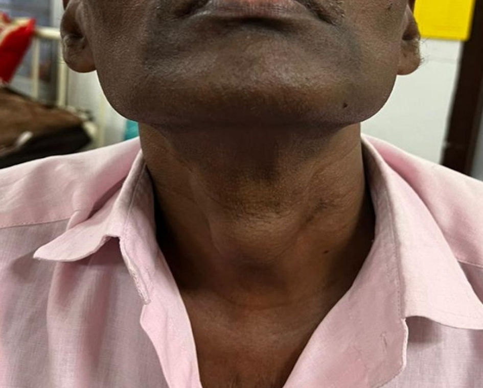 Click for large image | Figure 1. Midline swelling of thyroid gland. |
An anterior and lateral view radiographs of the cervical region revealed enhanced soft tissue opaqueness in the anterior neck area, without calcifications. USG of neck suggested a clearly defined isoechoic solid lesion in the center supra-thyroid region, indicating an ectopic or tensely filled persistent thyroglossal cyst.
CECT neck scan was suggestive of a clearly defined hypodense lesion in the midline, which is continuous into the superior margin of the thyroid isthmus. The possibilities include thyroglossal duct cancer, parathyroid adenoma, and infected thyroglossal duct cysts.
Positron emission tomography (PET) suggested enlargement in the center of the neck, superior to the thyroid gland, measuring 2.7 × 2.1 × 2.1 cm (Fig. 2). The tumor abutted the wind pipe and strap muscles with a probability of thyroglossal cyst neoplasia or paraganglioma.
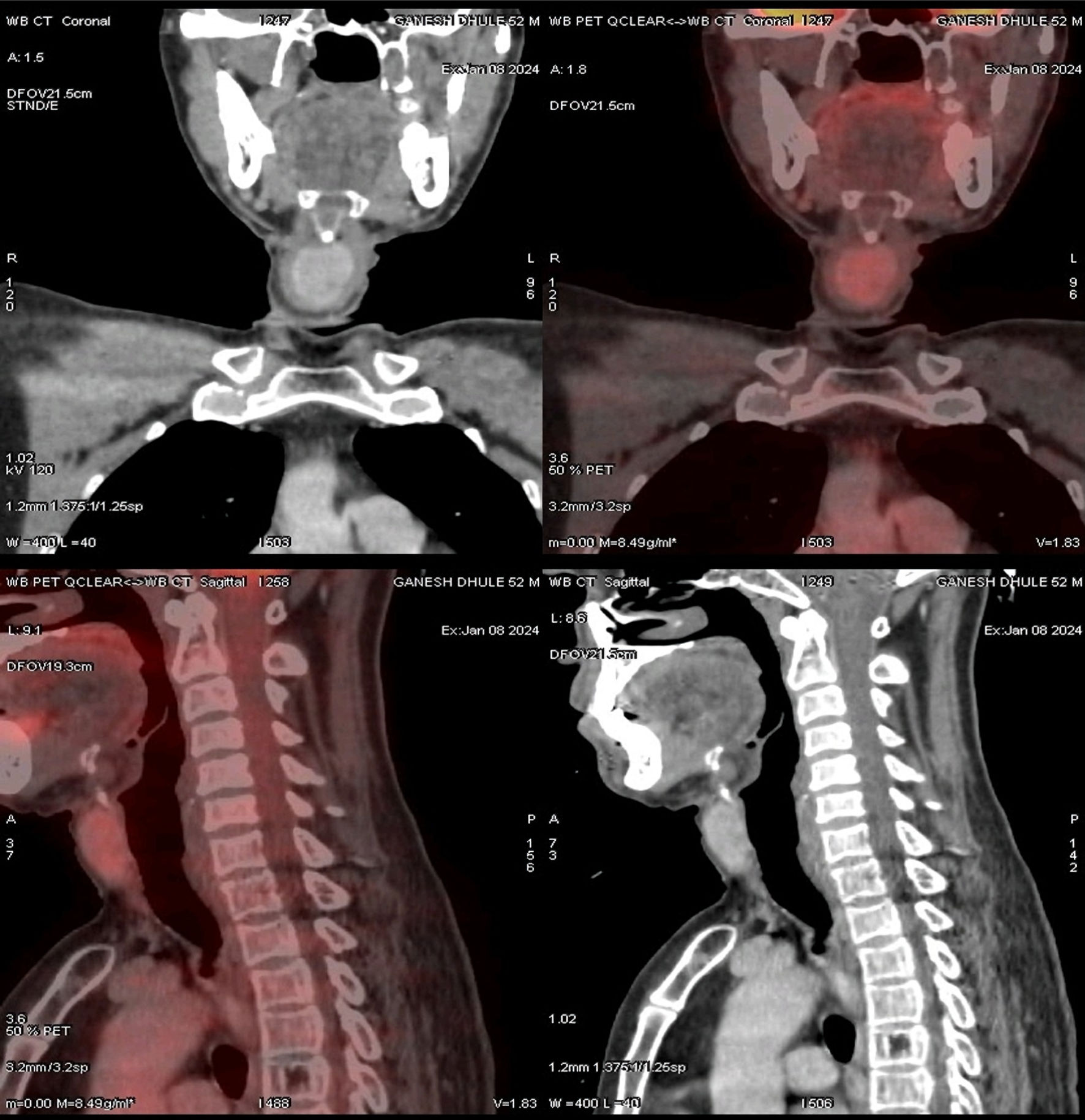 Click for large image | Figure 2. Computed tomography (CT) and positron emission tomography–computed tomography (PET-CT) demonstrating enlargement of the central portion of the thyroid gland. |
Test findings indicated serum calcitonin of 297 pg/mL, serum parathyroid hormone levels of 58.81 pg/mL, and free metanephrine of 5.67 pg/mL. There were no mutations discovered while testing for RET exons 10, 11, 13, 14, 15, and 16.
Fine-needle aspiration cytology (FNAC) revealed tumor cells that were either oval or round in shape, with moderate to widespread granular eosinophilic cytoplasm. The cell’s nucleus was circular to oval, having stippled (salt and pepper-like) chromatin. There was minimal to moderate nuclear and cellular morphological diversity. Papanicolaou staining (Pap) and Congo red stain results were inconclusive. The differential diagnoses included medullary thyroid cancer, parathyroid tumor, and paraganglioma.
The patient subsequently underwent complete thyroidectomy with bilateral neck dissection under general anesthesia. The specimen (Fig. 3) was sent for histological evaluation.
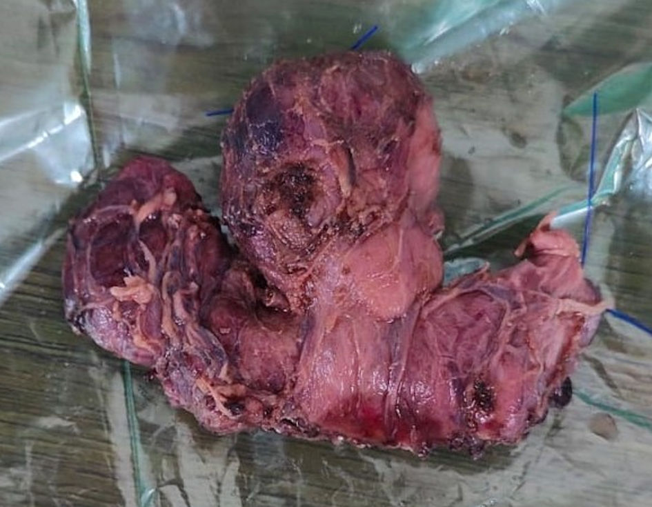 Click for large image | Figure 3. Total thyroidectomy specimen. |
Histopathology report indicated MTC, with the tumor epicenter located in the isthmus, with sizes of 2.8 × 2.5 × 1.5 cm and 0.4 × 0.4 × 0.3 cm, distributing throughout the isthmus and pyramidal lobe, encroaching towards both right and left lobe microscopically. There was a localized extrathyroidal spread with focal vascular embolization.
Immunohistochemistry results indicated positive staining of the tumor cells for calcitonin, CEA (Fig. 4), and thyroid transcription factor-1 (TTF1), but negative for paired box 8 (PAX8). The Ki-67 labelling index was 8–10%.
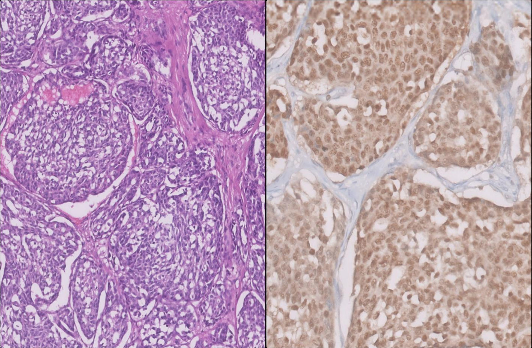 Click for large image | Figure 4. Histopathological and immunohistochemical images of medullary thyroid carcinoma (MTC) showing positivity for carcinoembryonic antigen (CEA). |
Two of the four nodes were metastatic. Lymph nodes located along the main specimen exhibited extranodal extension (dimension 0.1 cm).
Following surgery, USG revealed a 9 × 7 mm unevenly marginated hypoechoic lesion in the right thyroid bed and an 11 × 8 mm hypoechoic lesion in the suprasternal and pretracheal area.
Dodecane tetra acetic acid (DOTA) PET imaging suggested a low-grade somatostatin receptor (SSTR)-expressing pretracheal node, most likely a metastatic node. Repeat FNAC was predictive of metastatic medullary thyroid cancer.
The patient subsequently underwent revision surgery of right thyroid bed exploration combined with right cervical dissection and central compartment clearance.
The histopathology report indicated that there was no residual tumor in the right thyroid bed, and one of the 26 lymph nodes was positive for MTC. The positive lymph node corresponded to the central compartment.
Adjuvant external beam radiation therapy (EBRT) was administered to the tumor bed and draining nodes at a dosage of 60 Gy in 30 fractions over 6 weeks using the conformal approach.
The patient was monitored every 3 months after radiation therapy with cervical USG, calcitonin, and CEA levels. The results were within the normal range over 1-year follow-up.
| Discussion | ▴Top |
MTC usually appears in upper and middle thirds of both lobes. Tumors with the isthmus as the epicenter and extending across both lobes and the pyramidal lobe are rare. Almost all inherited MTC cases carry the RET proto-oncogene mutation, whereas such mutations are reported in as many as 50% of sporadic instances. Sporadic diseases lacking RET mutation may have RAS mutation.
MTC does not concentrate radioactive iodine particles and is not responsive to levothyroxine. It generally manifests as a unilateral isolated malignancy, whereas FMTC manifests as a bilateral multi-centric pattern. The MTC was restricted to one lobe in nearly 70% of the cases. Sporadic multifocal tumors with isthmus as epicenter spreading over both lobes are rare [4].
Serum calcitonin and CEA are key indicators for MTC and a whole-neck ultrasound is required to detect lymph nodes metastases. CECT, liver MRI, and bone scans are all needed in instances with calcitonin levels greater than 500 pg/mL to screen for distant metastases. If accessible, DOTA PET-CT can be explored for MTC patients with high blood calcitonin levels (≥ 500 pg/mL). In more advanced cases, somatic RET mutation testing may be necessary for selective RET inhibitor therapy [5].
In pre-surgically detected MTC, complete thyroidectomy is recommended due to the elevated frequency of multi-centric and bilateral illness, occurring in 15% and 5% of individuals with sporadic MTC localized to the neck, respectively. The recommended therapy is total removal of the tumor and associated lymph nodes. The American Thyroid Association (ATA) recommends central compartment node dissection (CND) and ipsilateral lymph node dissection (LND) for individuals with calcitonin levels over 20 ng/L, as well as preemptive contralateral LND for patients with circulating levels above 200 ng/L.
In patients with regionally advanced MTC, surgical intervention of the primary tumor and lymph node resection in the central and lateral neck compartments should employ a considerably less aggressive approach, to preserve optimal speech, swallowing function, parathyroid function, and shoulder movement,
Adult members of the family with probable MEN 2B syndrome who have a RET germline alteration should opt for preemptive total thyroidectomy including CND [6].
Comprehensive knowledge about the genetic mechanisms and immunological microenvironments associated with sporadic MTC has encouraged the advent of targeted approaches and immunotherapy. The simultaneous use of tyrosine kinase inhibitor (TKI)-targeted treatment, along with immune checkpoint inhibitors, appears to be a potential therapeutic strategy for managing patients with advanced MTC based upon specific tumor mutation profiles and tumor microenvironments [7].
ATA recommends considering adjuvant EBRT to the cervical area and mediastinum in patients at elevated risk for localized recurrences (residual macroscopic or microscopic illness extrathyroidal extension, or widespread lymphatic metastases), along with those susceptible to obstruction of the airway [8].
Serum calcitonin and CEA levels are analyzed 3 months after the operation to evaluate the effectiveness of the surgical procedure and direct subsequent treatment. Patients with insignificant calcitonin and CEA levels had excellent prognosis, with a 5-years resurgence of around 5% [6].
In the present case, the MTC originated from isthmus and pyramidal lobe of thyroid, i.e., exclusively from central part of thyroid and encroaching towards both lobes microscopically. All the oncological principles were followed in the treatment of the patient including multimodal approach and even genetic counselling and workup.
Revision surgery was also performed to achieve an R0 excision, followed by postoperative radiotherapy. Follow-up evaluations performed every 3 months, including serum calcitonin and CEA measurements and neck USG, were unremarkable throughout the 1-year follow-up period.
Conclusions
MTC is a comparatively less common but significant thyroid malignancy that typically requires a thorough diagnosis and vigorous treatment. This case emphasizes the significance of prompt identification using calcitonin levels and imaging, which leads to successful surgical therapy. Adjuvant therapeutics, such as EBRT, might further reduce recurrence risk, emphasizing the importance of ongoing surveillance and personalized treatment methods in MTC therapy.
Acknowledgments
The authors thank the Department of Pathology, Tata Hospital, for providing the histopathological and immunohistochemical images used in this report.
Financial Disclosure
None to declare.
Conflict of Interest
None to declare.
Informed Consent
Consent was obtained from the patient for publication of case report along with clinical images.
Author Contributions
Dr Gadi Venkatesh: Conception, design, data collection, analysis. Dr Kavita V. Jadhav: Conception, design, supervision, operating surgeon. Dr Rukmini P. Waghmare: Data processing, analysis, critical review. Dr Rajendra Habib: Review of literature, analysis, critical review. Dr Ritik M. Gandhi: Material and data collection. Dr Aditya Valavan: Data collection and workup of the patient.
Data Availability
Any inquiries regarding supporting data availability of this study should be directed to the corresponding author.
| References | ▴Top |
- Fugazzola L. Medullary thyroid cancer - an update. Best Pract Res Clin Endocrinol Metab. 2023;37(1):101655.
doi pubmed - Roy M, Chen H, Sippel RS. Current understanding and management of medullary thyroid cancer. Oncologist. 2013;18(10):1093-1100.
doi pubmed - Thomas CM, Asa SL, Ezzat S, Sawka AM, Goldstein D. Diagnosis and pathologic characteristics of medullary thyroid carcinoma-review of current guidelines. Curr Oncol. 2019;26(5):338-344.
doi pubmed - Hamdy O, Awny S, Metwally IH. Medullary thyroid cancer: epidemiological pattern and factors contributing to recurrence and metastasis. Ann R Coll Surg Engl. 2020;102(7):499-503.
doi pubmed - Kim M, Kim BH. Current guidelines for management of medullary thyroid carcinoma. Endocrinol Metab (Seoul). 2021;36(3):514-524.
doi pubmed - Jayasinghe R, Basnayake O, Jayarajah U, Seneviratne S. Management of medullary carcinoma of the thyroid: a review. J Int Med Res. 2022;50(7):3000605221110698.
doi pubmed - Bai Y, Niu D, Yao Q, Lin D, Kakudo K. Updates in the advances of sporadic medullary thyroid carcinoma: from the molecules to the clinic. Gland Surg. 2020;9(5):1847-1856.
doi pubmed - Bartz-Kurycki MA, Oluwo OE, Morris-Wiseman LF. Medullary thyroid carcinoma: recent advances in identification, treatment, and prognosis. Ther Adv Endocrinol Metab. 2021;12:20420188211049611.
doi pubmed
This article is distributed under the terms of the Creative Commons Attribution 4.0 International License (CC BY 4.0), which permits unrestricted use, distribution, and reproduction in any medium, including commercial use, provided the original work is properly cited.
Journal of Current Surgery is published by Elmer Press Inc.