| Journal of Current Surgery, ISSN 1927-1298 print, 1927-1301 online, Open Access |
| Article copyright, the authors; Journal compilation copyright, J Curr Surg and Elmer Press Inc |
| Journal website https://jcs.elmerpub.com |
Case Report
Volume 000, Number 000, August 2026, pages 000-000

Adult Splenic Flexure Colocolic Intussusception Due to a Submucosal Colonic Lipoma Presenting as Acute Abdomen
Muhammad Haris Latifa, Imran Khokharb, Maheen Afzalc, f, Eman Mazhard, Joel Rileye
aDepartment of Medicine, SSM Health St. Mary’s Hospital, St. Louis, MO, USA
bDepartment of Medicine, Reading Hospital Tower Health, West Reading, PA, USA
cDepartment of Medicine, University of Alabama at Birmingham, Montgomery, AL, USA
dDepartment of Medicine, Nishtar Medical University, Multan, Pakistan
eDepartment of Gastroenterology, SSM Health St. Mary’s Hospital, St. Louis, MO, USA
fCorresponding Author: Maheen Afzal, Department of Medicine, University of Alabama at Birmingham, Montgomery, USA
Manuscript submitted May 5, 2026, accepted June 18, 2026, published online August 7, 2026
Short title: Splenic Flexure Intussusception From Colonic Lipoma
doi: https://doi.org/10.14740/jcs1045
| Abstract | ▴Top |
Adult intussusception is uncommon and usually results from an underlying structural abnormality, particularly in the colon, where malignancy is a major concern. We present a case in which a young woman experienced 4 days of crampy lower abdominal pain, constipation, nausea, vomiting, and inability to pass flatus. Abdominal examination showed tenderness, hyperactive bowel sounds, and no distention; vital signs were stable. Laboratory tests were largely unremarkable, including a white blood cell count of 9.3 × 10⁹/L. Contrast-enhanced computed tomography of the abdomen identified a 9 cm colocolic intussusception at the splenic flexure, led by a 6 cm fat-containing lesion. Follow-up CT showed ongoing intussusception, bowel wall thickening, and a 7.2 × 3.8 cm fat-density lesion, confirming a fixed lead point. After a gastroenterology and surgical consultation, a laparoscopic left hemicolectomy was performed. Intraoperative findings confirmed intussusception, and histopathology revealed a benign submucosal colonic lipoma. This case underscores the rare occurrence of splenic flexure intussusception caused by a large submucosal lipoma, underscoring that persistent intussusception with a risk of malignancy warrants surgical management. Colonic lipoma should be considered in the differential diagnosis of adult colocolic intussusception, especially when malignancy cannot be excluded preoperatively.
Keywords: Adult intussusception; Colocolic intussusception; Splenic flexure; Colonic lipoma; Submucosal lipoma; Acute abdomen; Left hemicolectomy; Lead point lesion
| Introduction | ▴Top |
Adult intussusception is rare and represents a minor fraction of bowel obstruction cases compared to the pediatric population [1]. In contrast to childhood intussusception, which is generally idiopathic, adult cases are often associated with an identifiable structural lead point. This distinction is clinically significant, as neoplasia is prevalent in adult intussusception, especially when the colon is affected [2]. As a result, colonic intussusception in adults often raises concern for an underlying malignant lesion and typically necessitates definitive surgical resection rather than nonoperative reduction [3].
Colonic lipomas are benign mesenchymal tumors composed of mature adipose tissue, most commonly originating from the submucosa. Although many colonic lipomas are incidental and asymptomatic, larger lesions can result in abdominal pain, bleeding, altered bowel habits, obstruction, or serve as a lead point for intussusception [4]. In evaluating these cases, cross-sectional imaging is valuable, as colonic lipomas typically appear on computed tomography (CT) of the abdomen and pelvis as well-circumscribed intraluminal masses with fat attenuation. However, the presence of intussusception, infarction, or additional bowel wall changes may complicate radiologic interpretation [5, 6].
Adult colocolic intussusception caused by a benign colonic lipoma is rare, with splenic flexure involvement particularly uncommon. Because this presentation can closely resemble colonic malignancy both clinically and radiographically, it poses significant diagnostic and therapeutic challenges [7, 8]. To illustrate, this report describes a case of adult splenic-flexure colocolic intussusception caused by a large submucosal lipoma in a woman who presented with acute abdominal pain, nausea, vomiting, and obstipation, and was ultimately managed with left hemicolectomy.
| Case Report | ▴Top |
Investigations
A 34-year-old woman with a medical history of sinusitis, migraine, fibromyalgia, and gastroesophageal reflux disease presented with a 4-day history of crampy, nonradiating lower abdominal pain accompanied by constipation, nausea, nonbloody emesis, and inability to pass flatus. She reported no fever, weight loss, appetite changes, or recent medication adjustments. Upon presentation, vital signs were stable. Physical examination revealed a soft abdomen with mild lower abdominal tenderness and hypoactive bowel sounds, without evidence of peritoneal irritation.
Given her symptoms and examination findings, initial laboratory investigations were performed and were unremarkable. Results included a white blood cell count of 9.3 × 10⁹/L, hemoglobin of 11.3 g/dL, lactate of 0.9 mmol/L, normal renal function, lipase of 6 U/L, negative serum human chorionic gonadotropin, and an unremarkable urinalysis.
Diagnosis
To further evaluate her condition, CT of the abdomen and pelvis was obtained. This revealed a short-segment splenic flexure colocolic intussusception measuring approximately 9 cm, with a 6 cm fat-containing intraluminal lesion serving as the lead point, as illustrated in Figure 1. There was no evidence of lymphadenopathy, obstruction, perforation, or significant bowel dilatation. Repeat imaging the following day demonstrated persistent focal colonic intussusception, low-attenuation bowel wall thickening, and a fat-density intraluminal lesion measuring 7.2 × 3.8 cm, consistent with a fixed structural lead point, as illustrated in Figure 2.
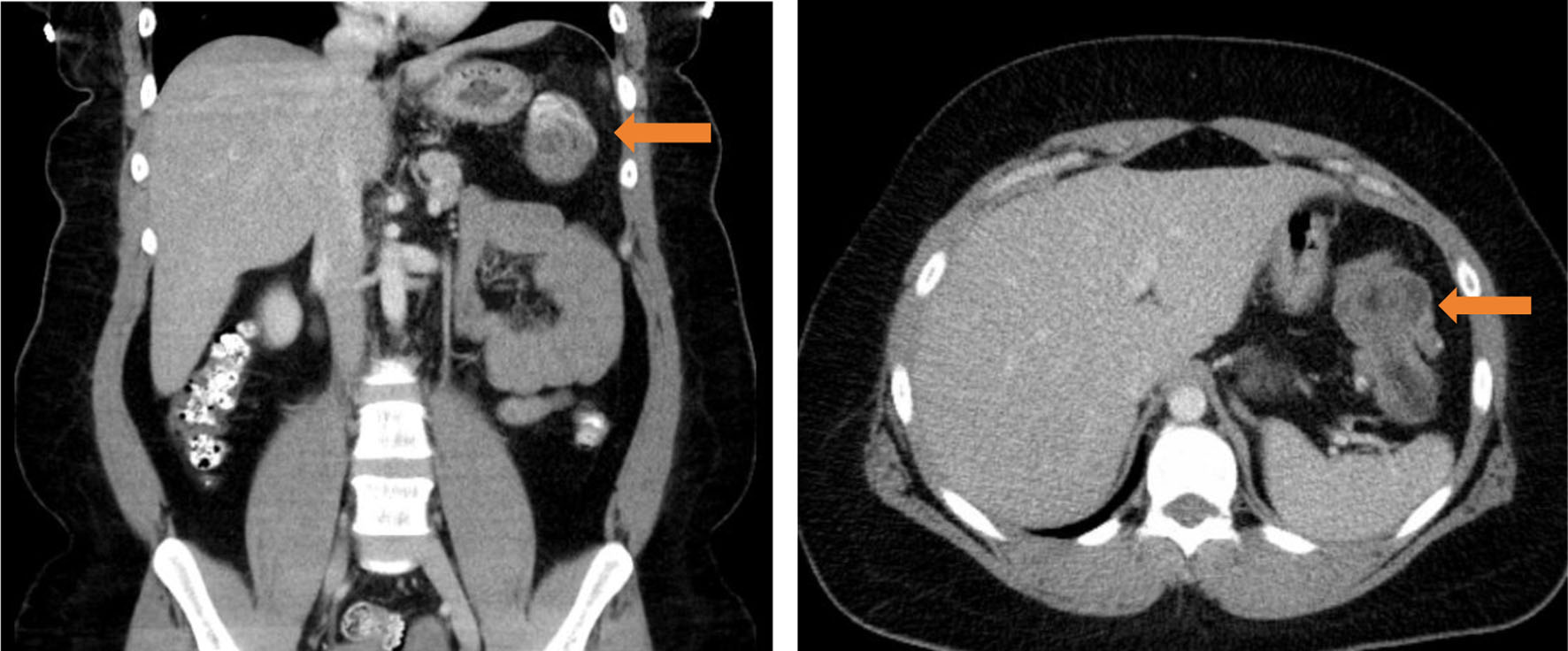 Click for large image | Figure 1. Initial computed tomography (CT) of abdomen/pelvis, coronal view (a), axial view (b), demonstrating splenic flexure colocolic intussusception with a lead point. |
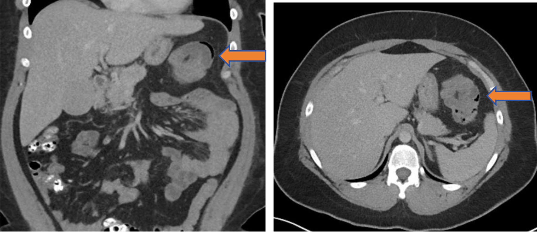 Click for large image | Figure 2. Follow-up computed tomography (CT) of abdomen/pelvis, coronal view (a) and axial view (b), demonstrating splenic flexure colocolic intussusception with a lead point indicated by blue arrows, showing low-attenuation bowel wall thickening with surrounding edema. |
Based on these imaging results, the primary diagnostic consideration was adult colocolic intussusception secondary to a lead point lesion. While imaging characteristics were suggestive of a lipoma, preoperative exclusion of malignancy was challenging due to the colonic location and persistent intussusception. The final diagnosis was adult splenic flexure colocolic intussusception caused by a submucosal colonic lipoma.
Treatment
Initial management consisted of intravenous fluids and analgesia. Subsequently, a gastroenterology consultation recommended definitive surgical intervention due to persistent adult colocolic intussusception and the presence of a substantial structural lead point lesion.
Accordingly, the patient underwent laparoscopic left hemicolectomy with mobilization of the splenic flexure and primary stapled anastomosis. Intraoperative findings confirmed colocolic intussusception at the splenic flexure, as demonstrated in Figure 3. The affected colonic segment was resected, perfusion was assessed using indocyanine green fluorescence imaging, and the specimen was submitted for permanent histopathologic evaluation. Gross examination of the resected specimen demonstrated an 8-cm pedunculated intraluminal colonic mass measuring 3.5–4 cm in diameter and partially obstructing the lumen. Sectioning revealed a yellow-tan fatty cut surface consistent with a lipoma.
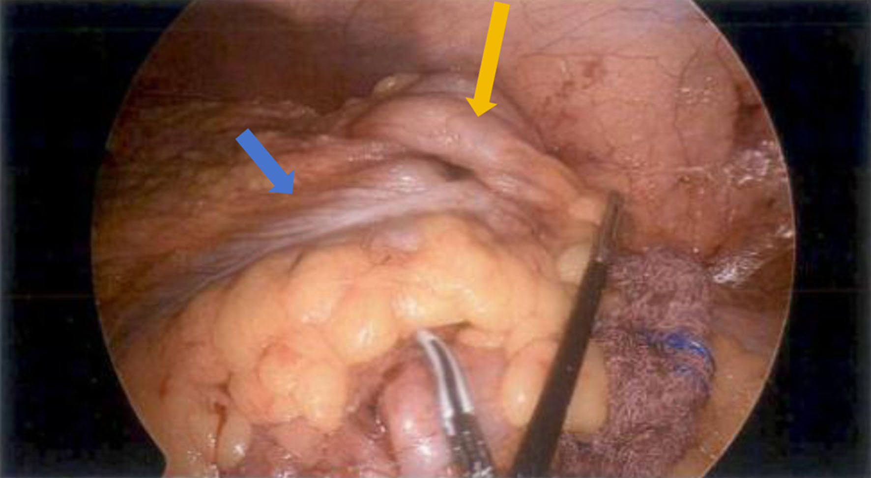 Click for large image | Figure 3. Intraoperative laparoscopic view of splenic flexure colocolic intussusception with a lead point lesion, as shown by the orange arrow with blue arrow showing the transverse colon. |
Follow-up and outcomes
The patient tolerated the procedure without complications, was extubated in the operating room, and transferred to the recovery unit in stable condition. Final histopathologic examination confirmed a benign submucosal colonic lipoma with no evidence of malignancy, as demonstrated in Figure 4.
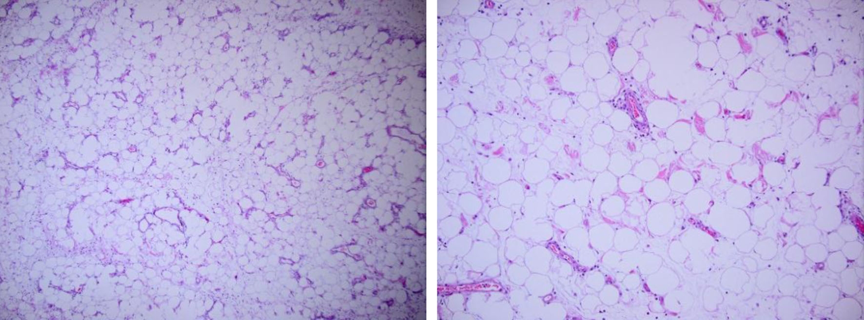 Click for large image | Figure 4. Histopathological examination of the lead point lesion demonstrating a submucosal colonic lipoma composed of mature adipocytes, confirming the benign nature of the intussusception lead point. |
The immediate postoperative course was favorable, with no intraoperative complications or unexpected adverse events documented.
Building on this favorable outcome, this case demonstrates a rare instance of adult splenic flexure colocolic intussusception caused by a benign submucosal lipoma, which initially raised suspicion for an underlying neoplastic lead point.
| Discussion | ▴Top |
This case demonstrates several key aspects of adult intussusception. Unlike pediatric cases, adult intussusception typically involves an underlying pathologic lead point. The likelihood of a structural lesion is especially high in colonic intussusception. Malignant neoplasms are a significant concern [2]. As a result, adult colonic intussusception is managed more aggressively than small-bowel intussusception. Oncologic resection without prior reduction is frequently recommended when malignancy cannot be confidently excluded [3]. In this case, persistent splenic flexure colocolic intussusception and a large, fixed lead-point lesion supported definitive resection.
This case demonstrates both the diagnostic value and limitations of CT in adult colocolic intussusception. The lead point was identified as a large, fat-containing intraluminal lesion at the splenic flexure. This finding was suggestive of a lipoma. The radiographic appearance is typical, as colonic lipomas generally show fat attenuation and smooth margins [9]. However, even if imaging favors a benign lipomatous lesion, conservative management is not appropriate for adult colonic intussusception. Bowel wall edema, intussusception-related distortion, and the malignant potential of colonic lead points may obscure the diagnosis. Previous imaging series show that intussuscepted lipomas may lose their characteristic homogeneous fat appearance. This can further complicate preoperative assessment [10–12].
The pathologic diagnosis in this case aligns with the established biology of colonic lipomas. These lesions are uncommon and typically submucosal. They often remain asymptomatic until they reach a significant size [13]. Larger lesions, especially those over 2 cm, are more likely to cause symptoms such as pain, bleeding, intermittent obstruction, or intussusception [14]. In this case, the lesion measured about 6–7 cm on serial imaging. This supports its role as the mechanical lead point for a 9 cm short-segment colocolic intussusception. Final pathology confirmed a submucosal lipoma without malignancy. This explains the benign lead point and highlights the difficulty of preoperative cancer exclusion.
The lipoma’s location at the splenic flexure underscores the educational significance of this report. Most documented cases of adult colonic lipoma with intussusception involve the ascending, transverse, or sigmoid colon, whereas splenic flexure presentations are distinctly rare [8, 15]. This anatomic site may complicate endoscopic or operative planning, particularly when symptoms progress over several days without overt perforation or complete obstruction. In this case, repeat imaging demonstrated persistent intussusception with low-attenuation bowel wall thickening, indicating a fixed lead point and possible bowel wall edema rather than a transient, self-limited event.
Management of lipoma depends on lesion size, symptoms, location, and diagnostic certainty. Small, incidentally detected lesions may be observed, and selected pedunculated or accessible lesions can be removed endoscopically [16]. In contrast, large symptomatic lesions causing intussusception generally require surgery, particularly when the lesion is broad-based, the diagnosis is uncertain, or the involved segment is in the colon, where malignancy risk is higher [17]. Our patient’s persistent symptoms, failure of spontaneous resolution, and imaging evidence of a large structural lead point supported surgical resection. Laparoscopic left hemicolectomy with primary anastomosis provided both definitive treatment and diagnostic confirmation.
Although surgical resection remains the standard treatment for adult colocolic intussusception, several reports have described successful endoscopic management of lipoma-associated intussusception in carefully selected patients. Orhan et al reported complete endoscopic treatment of ileocecal intussusception caused by a colonic lipoma without the need for surgery [18]. In another study, Schopis et al described successful endoscopic mucosal resection of massive colonic lipomas serving as lead points for intussusception [19]. Nevertheless, endoscopic reduction or resection should be reserved for highly selected patients with a strong preprocedural suspicion of benign disease, no evidence of bowel ischemia or perforation, and lesions that are technically amenable to endoscopic therapy [20]. In our patient, the large size of the lesion, persistent intussusception on serial imaging, and inability to confidently exclude malignancy favored definitive surgical resection rather than an attempt at endoscopic reduction.
This case should be considered within the context of the broader literature. The literature mostly consists of case reports, small series, and systematic reviews with heterogeneous reporting. As a result, estimates of malignancy risk and the optimal approach to reduction versus upfront resection vary across studies. However, the practical message remains the same: adult colocolic intussusception requires careful evaluation for an underlying organic lesion. Surgical management is often appropriate when a fixed colonic lead point is identified.
Key learning points
In summary, adult splenic flexure colocolic intussusception caused by a submucosal lipoma is rare but clinically significant. Even when CT imaging suggests a benign fat-containing lesion, persistent adult colonic intussusception should prompt strong consideration of definitive surgical resection, as malignancy cannot be reliably excluded preoperatively. This case underscores the importance of recognizing colonic lipoma as a benign but uncommon lead point for adult intussusception and highlights the rationale for operative management in this context.
Acknowledgments
The authors thank the clinical and surgical teams involved in the patient’s care. The authors also acknowledge the pathology and radiology departments for their contribution to diagnostic evaluation and confirmation of the final diagnosis.
Financial Disclosure
The authors received no financial support for the research, authorship, and/or publication of this article.
Conflict of Interest
The authors declare that they have no conflict of interest relevant to this manuscript.
Informed Consent
Written informed consent was obtained from the patient for publication of this case report and any accompanying images.
Author Contributions
Muhammad Haris Latif contributed to case identification, literature review, manuscript drafting, and final manuscript preparation. Imran Khokhar and Joel Riley contributed to the patient’s clinical management, the critical revision of the manuscript, and supervision. Maheen Afzal and Eman Mazhar contributed to data collection, interpretation of clinical findings, and manuscript editing. All authors reviewed and approved the final manuscript.
Data Availability
All data generated or analyzed during this study are included in this published article. Additional de-identified details are available from the corresponding author on reasonable request, subject to patient privacy considerations.
| References | ▴Top |
- Negametzyanov M, Smith CR, Ypsilantis E. Ileo-colic intussusception: a rare cause of intestinal obstruction in adults. J Surg Case Rep. 2025;2025(9):rjaf723.
doi pubmed - Marsicovetere P, Ivatury SJ, White B, Holubar SD. Intestinal Intussusception: Etiology, Diagnosis, and Treatment. Clin Colon Rectal Surg. 2017;30(1):30-39.
doi pubmed - Heersche S, Hirt J, Butti F, Hubner M, Hahnloser D, Joliat GR, Grass F. Intestinal Intussusception in Adults: A Systematic Review. World J Surg. 2025;49(10):2706-2716.
doi pubmed - Habte YM, Habte BM, Kifle YA, Abdu EM, Yusuf YM, Yimer SA. Large colonic lipomas presenting as a rare cause of adult bowel obstruction and intussusception: A two-case surgical series. Int J Surg Case Rep. 2025;136:111988.
doi pubmed - Fiordaliso M, Lovaglio UM, De Marco FA, Costantini R, Nasti GA, Lelli Chiesa P. Colonic lipoma, a rare cause of intestinal intussusception: A narrative review and how to diagnose it. Medicine (Baltimore). 2024;103(39):e39579.
doi pubmed - Buetow PC, Buck JL, Carr NJ, Pantongrag-Brown L, Ros PR, Cruess DF. Intussuscepted colonic lipomas: loss of fat attenuation on CT with pathologic correlation in 10 cases. Abdom Imaging. 1996;21(2):153-156.
doi pubmed - Moussally M, Mokalled I, Jamali F, Khalife MJ. Splenic flexure colonic lipoma causing intussusception. JRSM Open. 2021;12(1):2054270420983088.
doi pubmed - Acharya A, Acharya A, Chaulagain U, Khanal K, Shah S. Colocolic intussusception caused by a descending colon lipoma in an adult: a rare case report. Int J Surg Case Rep. 2026;138(4):1287-1292.
doi pubmed - Bashir S, Bhat GA, Nazir Y, Wagay BA, Lone ZG, Malik AA. Giant colonic lipoma causing intussusception: a rare case report and literature review. World J Colorectal Surg. 2025;14(4):132-135.
doi - Kim YH, Blake MA, Harisinghani MG, Archer-Arroyo K, Hahn PF, Pitman MB, Mueller PR. Adult intestinal intussusception: CT appearances and identification of a causative lead point. Radiographics. 2006;26(3):733-744.
doi pubmed - Gayer G, Hertz M, Zissin R. CT findings of intussusception in adults. Semin Ultrasound CT MR. 2003;24(5):377-386.
doi pubmed - Marinis A, Yiallourou A, Samanides L, Dafnios N, Anastasopoulos G, Vassiliou I, Theodosopoulos T. Intussusception of the bowel in adults: a review. World J Gastroenterol. 2009;15(4):407-411.
doi pubmed - Uygur FA, Kuloglu E, Aydin G, Muhtaroglu A, Dulger AC. Characterizing colon lipomas: Insights from a retrospective analysis of clinical presentation and management strategies. Medicine (Baltimore). 2024;103(21):e38287.
doi pubmed - An HH, Duong TT, Van Truong N, Van Quoc L, Son VN, Thang NP, Van Thach N, et al. A large lipoma of the descending colon: A rare case report. Radiol Case Rep. 2021;16(11):3396-3399.
doi pubmed - Menegon Tasselli F, Urraro F, Sciaudone G, Bagaglini G, Pagliuca F, Reginelli A, Ferraraccio F, et al. Colonic lipoma causing bowel intussusception: an up-to-date systematic review. J Clin Med. 2021;10(21):5149.
doi pubmed - Lee CS, Lee MJ, Kim KL, Kim YS, Baik GH, Kim JB, Kim DJ, et al. A case of giant lipoma causing chronic recurrent intussusception of the colon. Clin Endosc. 2012;45(2):165-168.
doi pubmed - Erginoz E, Uludag SS, Cavus GH, Zengin K, Ozcelik MF. Clinicopathological features and management of colonic lipomas: Case reports. Medicine (Baltimore). 2022;101(10):e29004.
doi pubmed - Orhan A, Demiryas S. Lipoma causing ileocecal intussusception and its endoscopic resection. Int J Surg Case Rep. 2022;98:107605.
doi pubmed - Schopis M, Yang J. Endoscopic Treatment of Intussusception From Massive Colonic Lipomas via Endoscopic Mucosal Resection: A Case Series. ACG Case Rep J. 2019;6(9):e00177.
doi pubmed - Zhou S, Chen Z, Hu Y. Successful endoscopic reduction of rare post-ESD colonic intussusception: A case report. Sci Prog. 2025;108(3):368504251375713.
doi pubmed
This article is distributed under the terms of the Creative Commons Attribution 4.0 International License (CC BY 4.0), which permits unrestricted use, distribution, and reproduction in any medium, including commercial use, provided the original work is properly cited.
Journal of Current Surgery is published by Elmer Press Inc.