Chronic Non-Infected Branchial Cleft Cyst With Jugular Vein Displacement: Diagnostic and Surgical Challenges in a Rare Neck Mass
DOI:
https://doi.org/10.14740/jcs1004Keywords:
Branchial cleft cyst, Neck swelling, Second branchial cleft cyst, Neck massAbstract
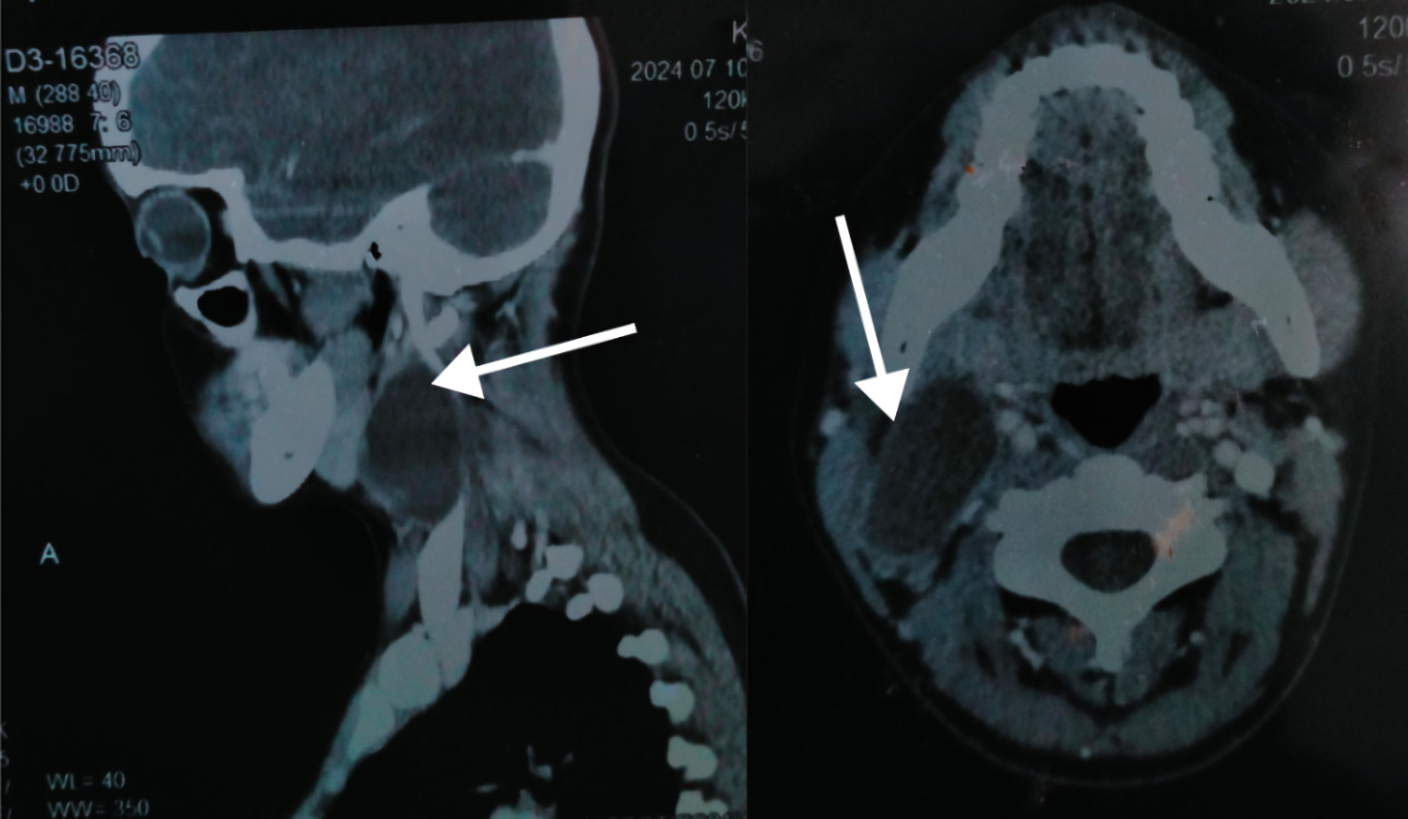
Branchial cleft anomalies are rare embryonic developmental abnormalities. We discuss a case of a 23-year-old female, who presented with a progressively enlarged swelling on the right side of her neck, which had been present since 2016 (8 years ago). Initially asymptomatic, the swelling progressively transformed, triggering intermittent pain during sore throat, and specific activities like loud speaking or long journeys. Multiple aspiration procedures offered temporary relief but failed to resolve the underlying pathology. The diagnostic challenge was profound, as it involved a rare condition mimicking more prevalent regional pathologies, such as tuberculosis and lymphadenopathy, particularly in Pakistan. Ultrasound, computed tomography (CT) scan and magnetic resonance imaging (MRI) meticulously mapped the lesion’s intricate anatomical relationships. These diagnostic modalities strongly suggested a second branchial cleft cyst, subsequently confirmed through histopathological examination. Surgical intervention involved complete cyst excision performed under general anesthesia. Microscopic analysis revealed a unilocular cyst with characteristic stratified squamous epithelial lining and associated lymphoid tissue. Critically, no malignant transformations were detected, with accompanying lymph nodes demonstrating benign reactive changes. This case is unique due to jugular vein displacement due to branchial cyst and the absence of infection over an extended period, from 2016 to 2024, despite the cyst’s tendency for infection as reported in the literature. It highlights the importance of early diagnosis, advanced imaging, and complete surgical excision to achieve favorable outcomes. Learning points include recognizing branchial cleft cysts in the differential diagnosis of lateral neck masses, the essential role of imaging in diagnosis and surgical planning, and the significance of histopathology in confirming the benign nature of such anomalies.
Published
Issue
Section
License
Copyright (c) 2025 The authors

This work is licensed under a Creative Commons Attribution-NonCommercial 4.0 International License.