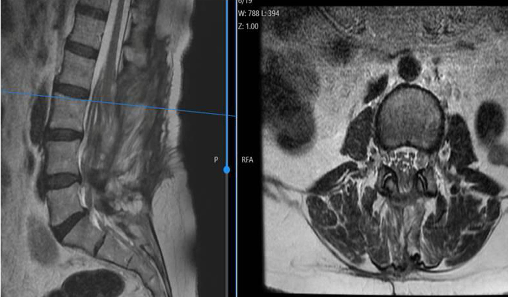
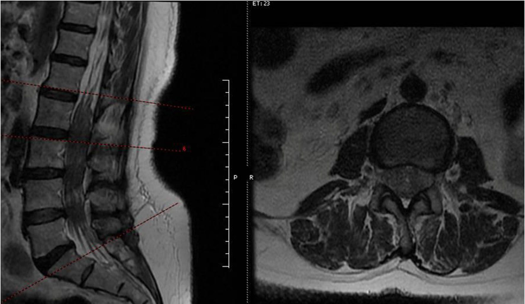
↓ Figure 1. Preoperative T2-weighted lumbar MRI showing diffuse hypointense intradural appearance extending from the conus medullaris to the L4–L5 level. MRI: magnetic resonance imaging.
| Journal of Current Surgery, ISSN 1927-1298 print, 1927-1301 online, Open Access |
| Article copyright, the authors; Journal compilation copyright, J Curr Surg and Elmer Press Inc |
| Journal website https://jcs.elmerpub.com |
Case Report
Volume 16, Number 2, September 2026, pages 54-58
Idiopathic Spinal Arachnoiditis Presenting With Acute Bilateral Lower Extremity Weakness
Figures