Figures
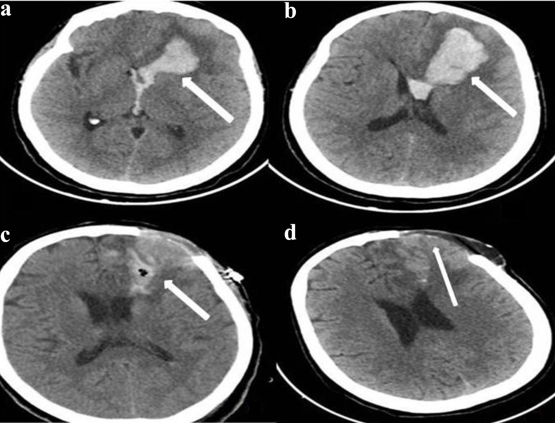
↓ Figure 1. Axial brain computed tomography (CT) sections with a slice thickness of 2.5 mm. Images (a) and (b) are preoperative scans demonstrating hyperdense hemorrhagic areas in the left frontal region with extension into the ventricle (the white arrow indicates the hemorrhage). Image (c) is a 2.5-mm axial CT scan obtained in the early postoperative period, 3 days after surgery (the white arrow indicates early postoperative changes at the surgical site). Image (d) is a 2.5-mm axial CT scan obtained 1 month after surgery, showing no evidence of hemorrhage (the white arrow indicates the previous hemorrhage evacuation site and the bone defect area).
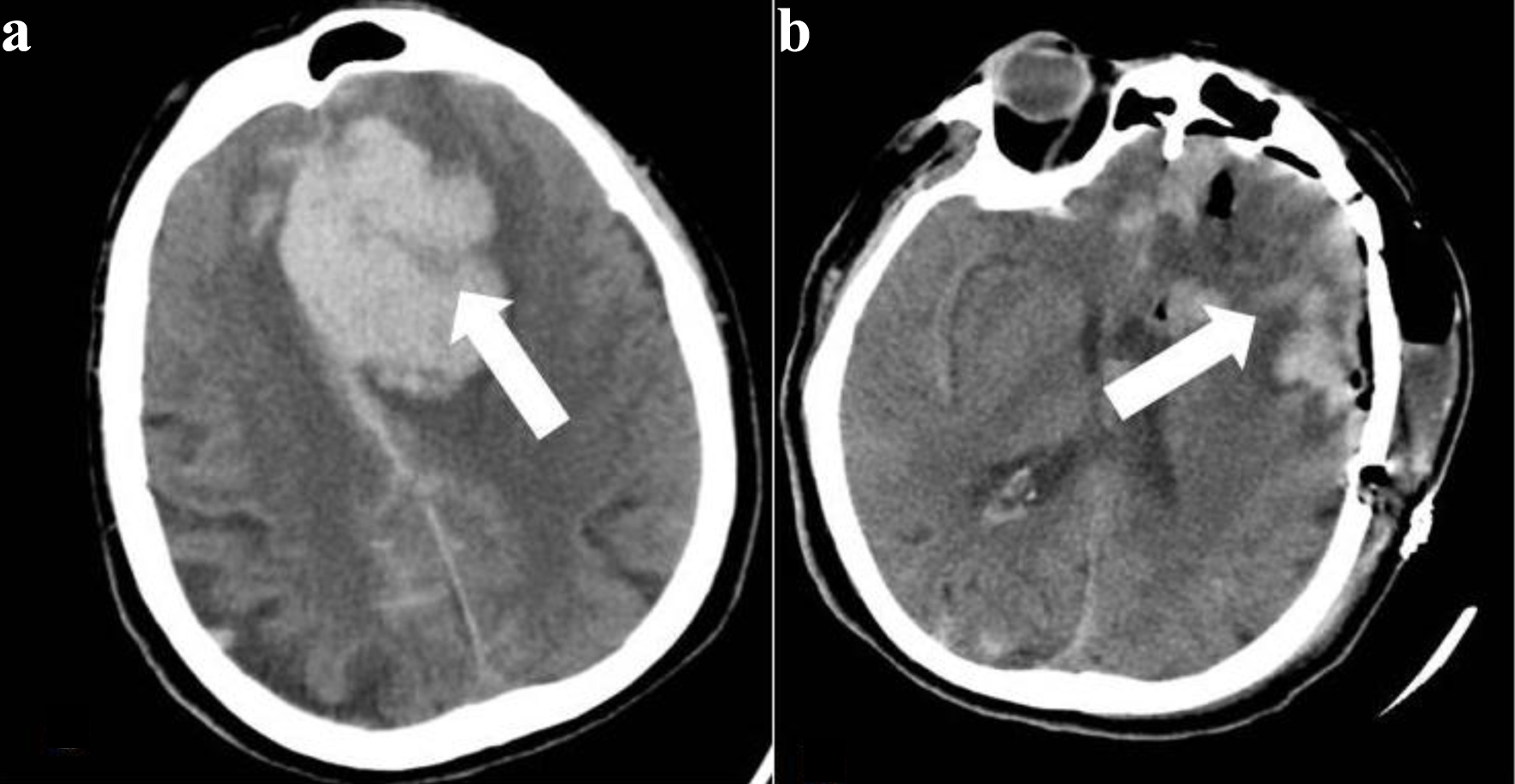
↓ Figure 2. Axial brain computed tomography (CT) sections with a slice thickness of 2.5 mm. (a) The preoperative image demonstrates a hematoma in the left frontal region causing a pronounced midline shift (the white arrow indicates the hemorrhagic area). (b) The 2.5-mm axial brain CT obtained 12 h after surgery shows findings consistent with craniotomy, with a marked regression of the midline shift (the white arrow indicates the surgical site, and the bone flap has been replaced).
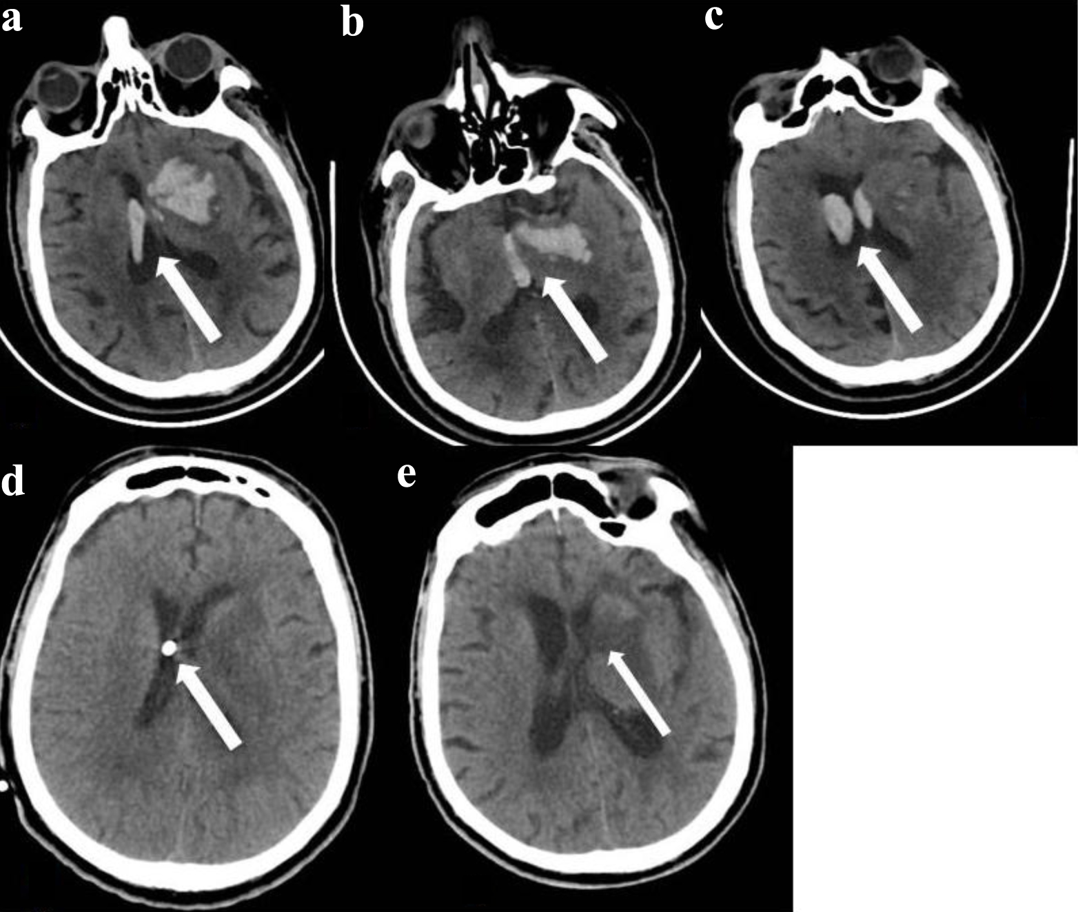
↓ Figure 3. Axial brain computed tomography (CT) sections with a slice thickness of 2.5 mm. (a) Hemorrhage is present in the right lateral ventricle and in the left lateral ventricle with parenchymal involvement (the white arrow indicates the hemorrhage). (b) Hemorrhage is observed in the left lateral ventricle, parenchyma, and the third ventricle (the white arrow indicates the hemorrhage). (c) Hemorrhage is present in both ventricles (the white arrow indicates the hemorrhage). (d) A 2.5-mm axial brain CT image obtained after placement of an external ventricular drainage (EVD) system shows no evidence of intraventricular hemorrhage (the white arrow indicates the ventricular tip of the EVD). (e) Axial brain CT images obtained 25 days after surgery demonstrate complete resorption of the hematoma. The EVD has been removed. An area corresponding to resorption of the parenchymal hematoma is seen in the left frontal region (the white arrow indicates the area of the resorbed parenchymal hematoma).
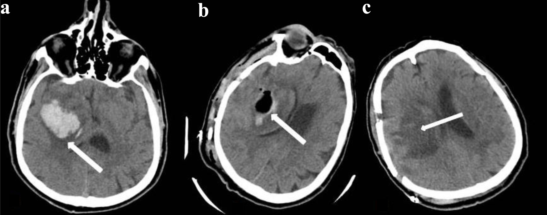
↓ Figure 4. Axial brain computed tomography (CT) sections with a slice thickness of 2.5 mm. (a) A hematoma is present in the right temporal region causing a midline shift (the white arrow indicates the hemorrhage). (b) An axial brain CT obtained 12 h after surgery demonstrates early postoperative changes with regression of the midline shift. The bone flap has been replaced (the white arrow indicates the evacuated hematoma cavity). (c) A 2.5-mm axial brain CT obtained 1 month after surgery shows complete resolution of the hematoma. Bone defects related to the craniotomy are present (the white arrow indicates the previously evacuated hematoma area).