Figures
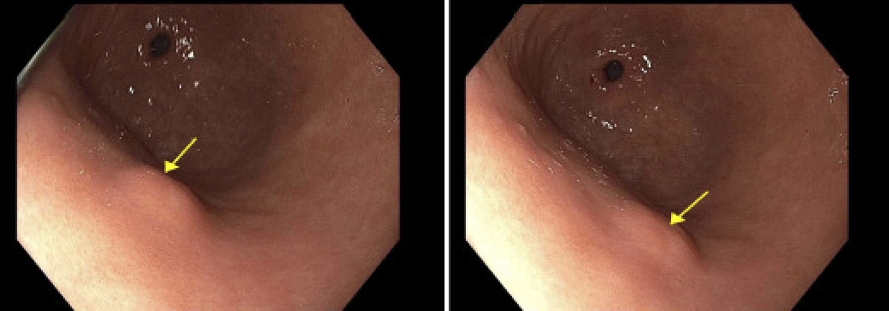
↓ Figure 1. Endoscopic image showing a single 8
mm submucosal nodule (arrows) on the anterior wall of the gastric body. The lesion appeared hyperechoic
on endoscopic ultrasound and invaded into the submucosa (layer 3).
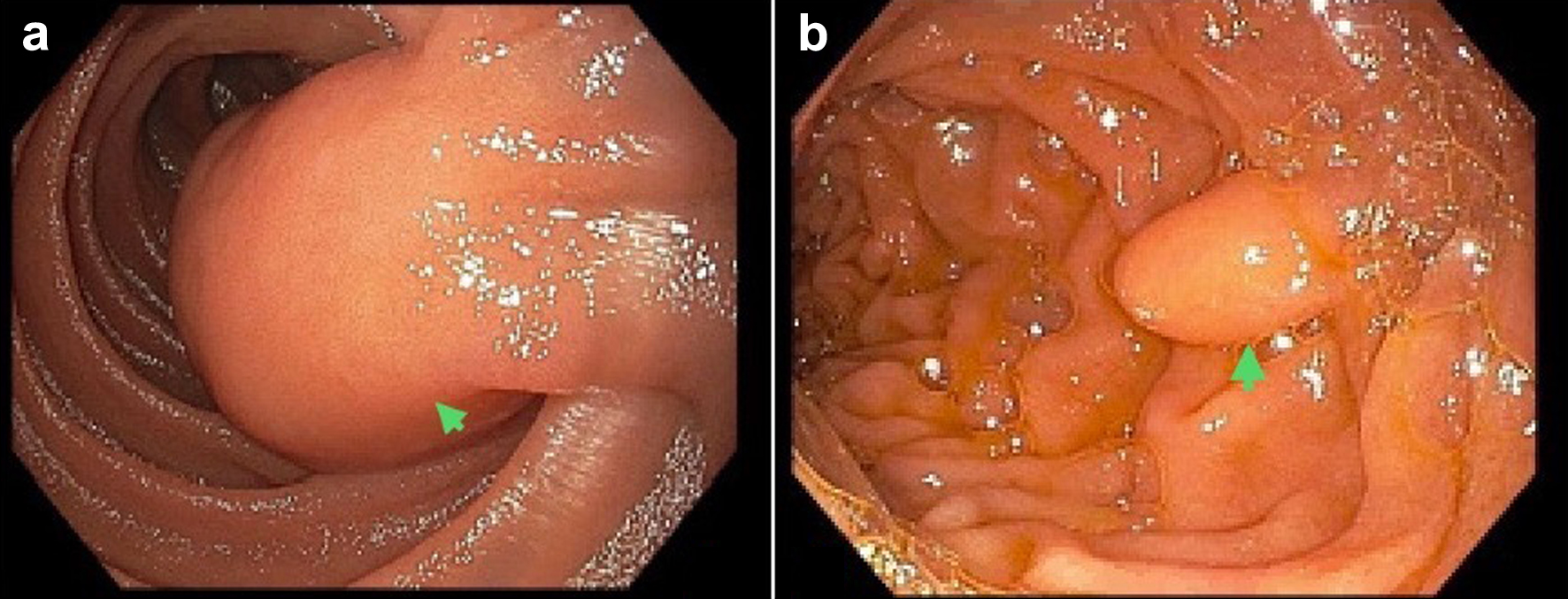
↓ Figure 2. Endoscopic images illustrating
lipomas in the small intestines. (a) A mass in the third portion of the duodenum (arrow). Histopathology
was consistent with a duodenal lipoma. (b) A medium-sized lipoma, measuring 12 mm in diameter, in the
second portion of the duodenum (arrow).
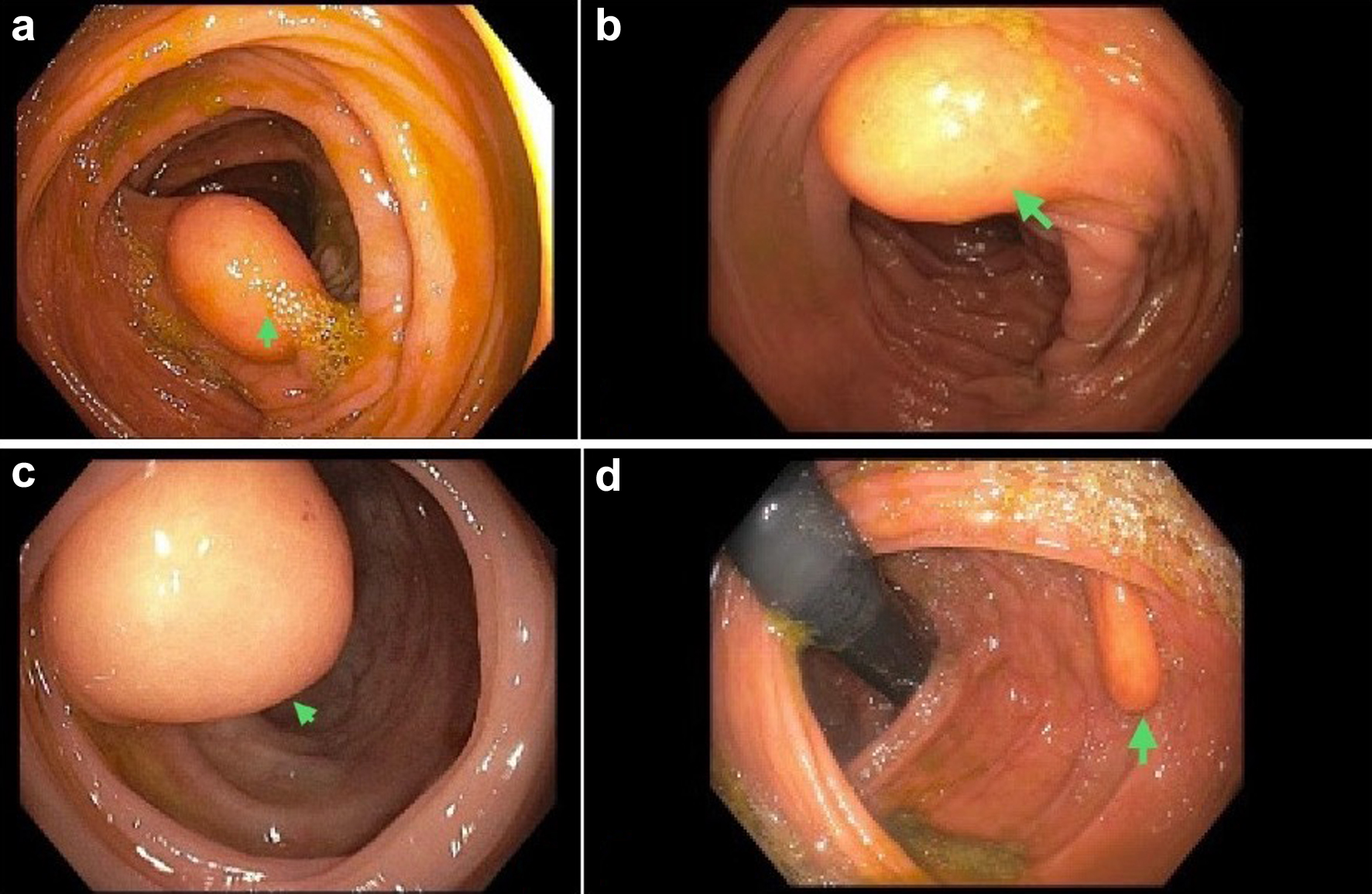
↓ Figure 3. A library of endoscopic images
showing colorectal lipomas. (a) A large lipoma, measuring 15 mm in diameter, in the ascending colon. (b)
A medium-sized lipoma at the hepatic flexure. (c) A large lipoma at the ileocecal valve. (d) A
medium-sized lipoma at the splenic flexure, in the transverse and ascending colon.
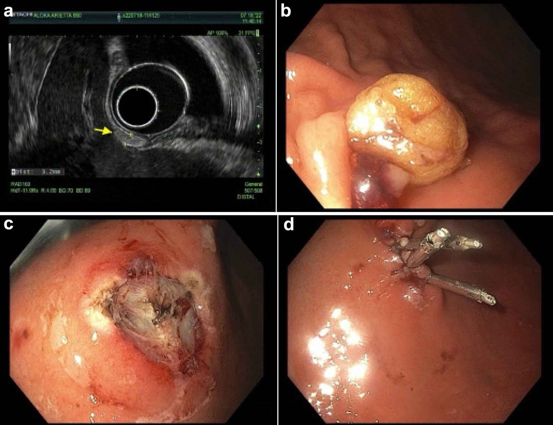
↓ Figure 4. A library of endoscopy images showing
the diagnosis and endoscopic mucosal resection of a gastric lipoma. (a) A hyperechoic oval nodule,
measuring 3.2 mm in maximal cross-sectional diameter. There was sonographic evidence suggesting invasion
into the submucosa. (b, c) A mucosectomy scar after mucosal resection of the lipoma. Three hemostatic
clips were employed to close the surgical mucosal defect (d).