| Journal of Current Surgery, ISSN 1927-1298 print, 1927-1301 online, Open Access |
| Article copyright, the authors; Journal compilation copyright, J Curr Surg and Elmer Press Inc |
| Journal website https://jcs.elmerpub.com |
Case Report
Volume 16, Number 2, September 2026, pages 55-59

Idiopathic Spinal Arachnoiditis Presenting With Acute Bilateral Lower Extremity Weakness
Hakan Aka, c  , Fatih Durnab , Sedanur Ozgulb , Emine Avginb
aDepartment of Neurosurgery, Kirsehir Ahi Evran University School of Medicine, Kirsehir, Turkiye
bKirsehir Training and Research Hospital, Kirsehir, Turkiye
cCorresponding Author: Hakan Ak, Department of Neurosurgery, Kirsehir Ahi Evran University School of Medicine, Turkiye
Manuscript submitted February 2, 2026, accepted July 15, 2026, published online August 29, 2026
Short title: Spinal Arachnoiditis
doi: https://doi.org/10.14740/jcs1027
| Abstract | ▴Top |
Arachnoiditis is a rare inflammatory disorder of the arachnoid membrane that may result in progressive neurological deficits. Due to its nonspecific clinical presentation and variable radiological findings, diagnosis remains challenging. A 61-year-old female presented with a 1-week history of progressive numbness and weakness in the bilateral distal lower extremities, resulting in inability to ambulate. Neurological examination revealed bilateral hypoesthesia below the L1 level and severe motor weakness. Lumbar magnetic resonance imaging demonstrated a diffuse intradural hypointense appearance on T2-weighted images extending from the conus medullaris to the L4–L5 level without contrast enhancement. Surgical exploration was performed for diagnostic and therapeutic purposes. No intradural mass lesion was identified; however, marked nerve root edema and extensive arachnoid adhesions were observed. Adhesiolysis and duraplasty were performed. Postoperatively, the patient showed significant neurological improvement. Idiopathic arachnoiditis should be considered in the differential diagnosis of acute or subacute bilateral lower extremity weakness. Surgical intervention may be beneficial in selected patients for both diagnosis and symptom relief, even when radiological findings persist.
Keywords: Arachnoiditis; Idiopathic; Spinal cord; Magnetic resonance imaging
| Introduction | ▴Top |
Arachnoiditis is a rare inflammatory disorder of the arachnoid membrane that can lead to significant neurological morbidity [1]. The condition has been associated with various etiological factors, including trauma, infection, iatrogenic injury, aneurysms, and neoplastic processes. In a subset of patients, no identifiable cause is found, and the condition is classified as idiopathic arachnoiditis [1–4].
Histopathologically, arachnoiditis progresses through three stages. The early stage is characterized by inflammation-induced hyperemia and edema of the nerve roots. This is followed by a proliferative stage marked by fibroblast activation and collagen deposition, leading to the formation of adhesions. In the advanced stage, progressive fibrosis results in atrophy of the affected nerve roots [5, 6].
Clinical presentation varies depending on the anatomical location and stage of the disease. Patients may present with localized pain, sensory disturbances of the extremities, motor weakness, muscle cramps, and autonomic dysfunction, including urinary or fecal incontinence and fatigue [1, 3]. Due to its nonspecific symptoms and resemblance to other spinal or intracranial pathologies, arachnoiditis poses a diagnostic challenge and requires meticulous clinical assessment and differential diagnosis [4].
Magnetic resonance imaging (MRI) is considered the gold standard for the diagnosis of arachnoiditis [1, 3, 4]. MRI findings may reveal the underlying cause, most commonly arachnoid cysts. Additional characteristic findings include dorsal or ventral tethering of the spinal cord due to adhesions, clumping of nerve roots producing a pseudocord appearance, and abnormal spinal cord dimensions related to edema and inflammation [6].
Management strategies for arachnoiditis are determined by disease severity and clinical progression. Conservative treatment is generally recommended in mild cases, whereas invasive interventions and surgical procedures may be required in advanced or refractory cases [1].
Herein, we present the case of a 61-year-old female patient who was admitted with a 1-week history of numbness and weakness in the bilateral distal lower extremities and was diagnosed with arachnoiditis.
| Case Report | ▴Top |
A 61-year-old female patient was admitted to our clinic with a 1-week history of progressive numbness and weakness in the bilateral distal lower extremities. Due to severe weakness, the patient was unable to ambulate. There was no evidence of sphincter dysfunction. The patient had no history of trauma, spinal surgery, or recent infection. Her medical history was significant for hypertension and rheumatoid arthritis. She was not receiving any medication other than amlodipine.
Neurological examination revealed normal cranial nerve and cerebellar functions. Muscle strength and sensory examination of the upper extremities were within normal limits. Sensory examination of the lower extremities demonstrated bilateral hypoesthesia below the L1 dermatome. Motor strength in the lower extremities was graded as 2/5 bilaterally. Deep tendon reflexes, including patellar and Achilles reflexes, were bilaterally decreased.
Laboratory investigations, including complete blood count, erythrocyte sedimentation rate, C-reactive protein, and routine biochemical parameters, were within normal ranges. Magnetic resonance imaging (MRI) of the brain, cervical spine, and thoracic spine revealed no pathological findings. Lumbar spine MRI demonstrated a hypointense lesion on T2-weighted images occupying the entire spinal canal, extending from the inferior margin of the conus medullaris to the L4–L5 level. Contrast-enhanced lumbar MRI showed no contrast enhancement (Figs. 1 and 2).
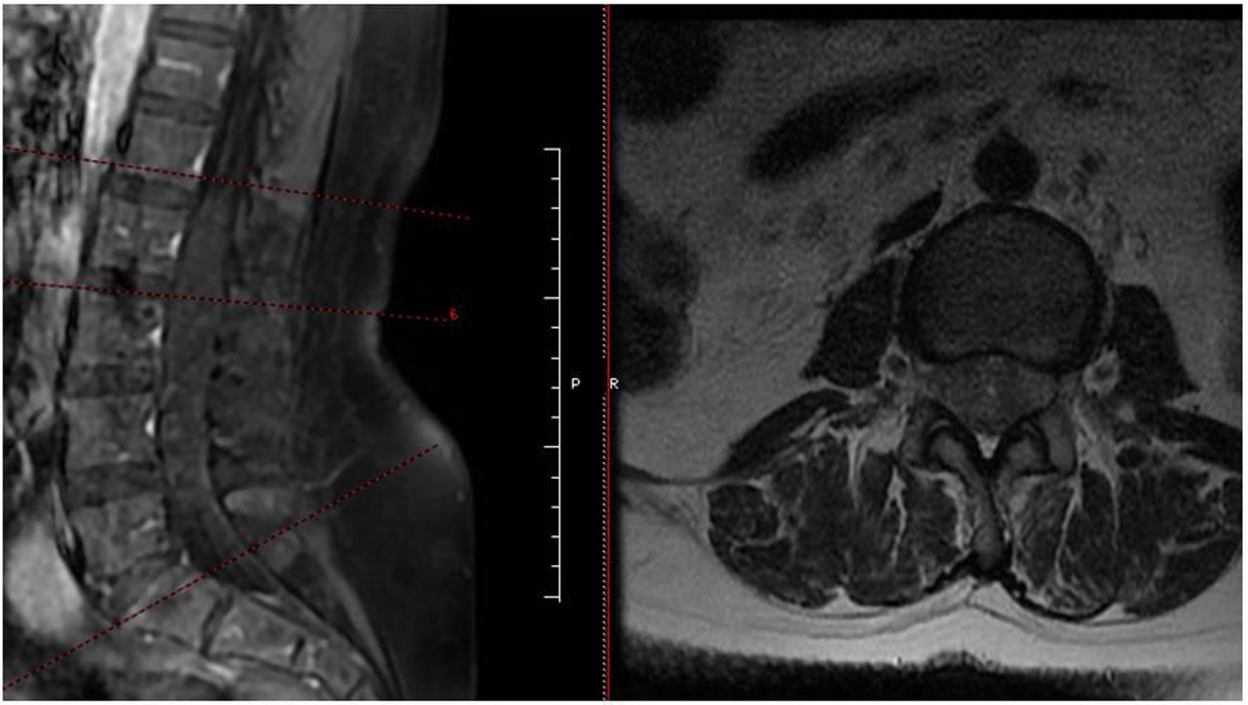 Click for large image | Figure 1. Preoperative T2-weighted lumbar MRI showing diffuse hypointense intradural appearance extending from the conus medullaris to the L4–L5 level. MRI: magnetic resonance imaging. |
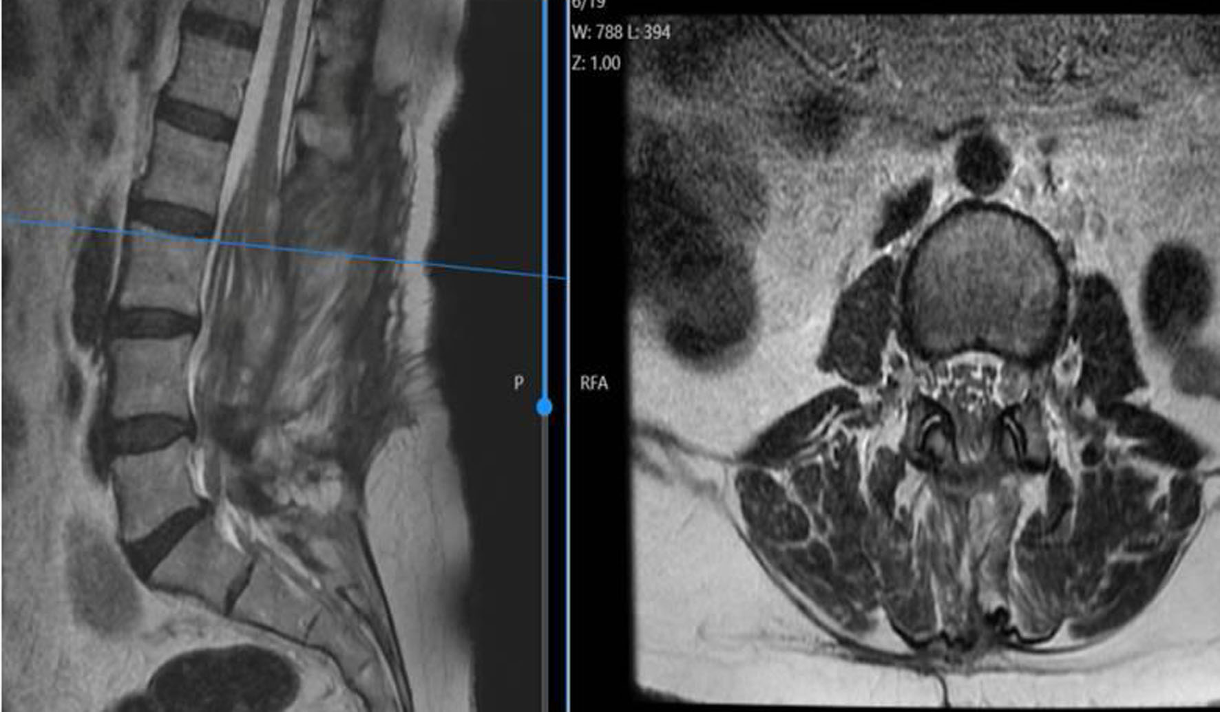 Click for large image | Figure 2. Contrast-enhanced lumbar MRI demonstrating absence of contrast enhancement. MRI: magnetic resonance imaging. |
Surgical intervention was undertaken for both diagnostic and therapeutic purposes. Following laminectomy, the dura mater was opened via a midline incision. No intradural mass lesion, such as tumor, abscess, or hematoma, was identified. However, all nerve rootlets were markedly swollen and edematous (Fig. 3). Arachnoid adhesions and fibrous bands were carefully dissected. Cerebrospinal fluid (CSF) samples were obtained for microbiological culture and cytological analysis. Duraplasty was subsequently performed using a fascia graft. CSF cultures showed no microbial growth, and cytological examination revealed no abnormal findings.
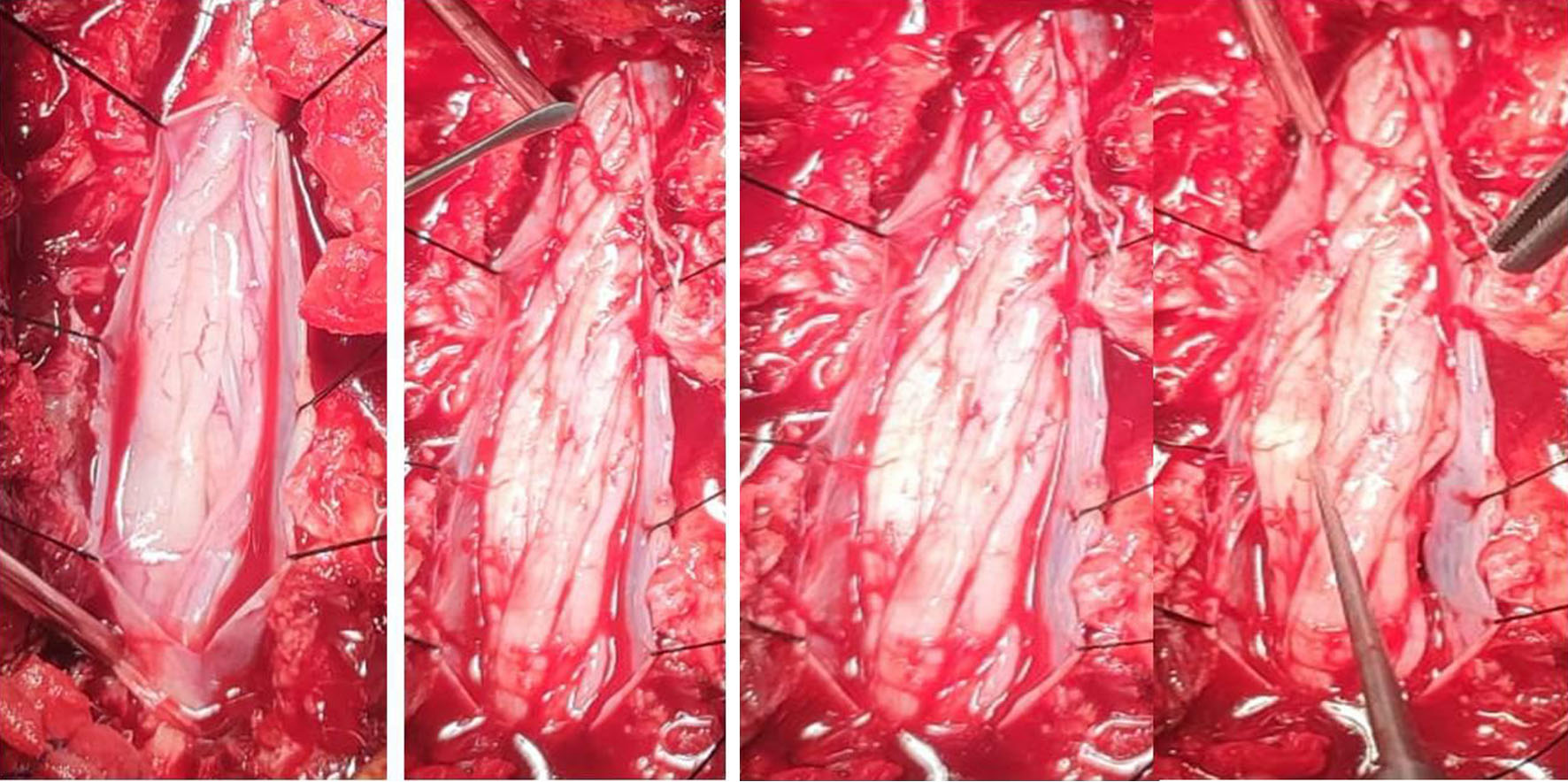 Click for large image | Figure 3. Intraoperative images showing markedly edematous and swollen nerve rootlets with arachnoid adhesions. |
At the 1-month follow-up, the patient demonstrated significant clinical improvement, with lower extremity muscle strength increasing to 4/5, and she was able to ambulate independently. A marked reduction in numbness was also noted. Follow-up lumbar MRI performed at the third and sixth months demonstrated partial radiological improvement, with decreased clumping and crowding of the nerve rootlets; however, the nerve roots remained swollen and edematous (Fig. 4).
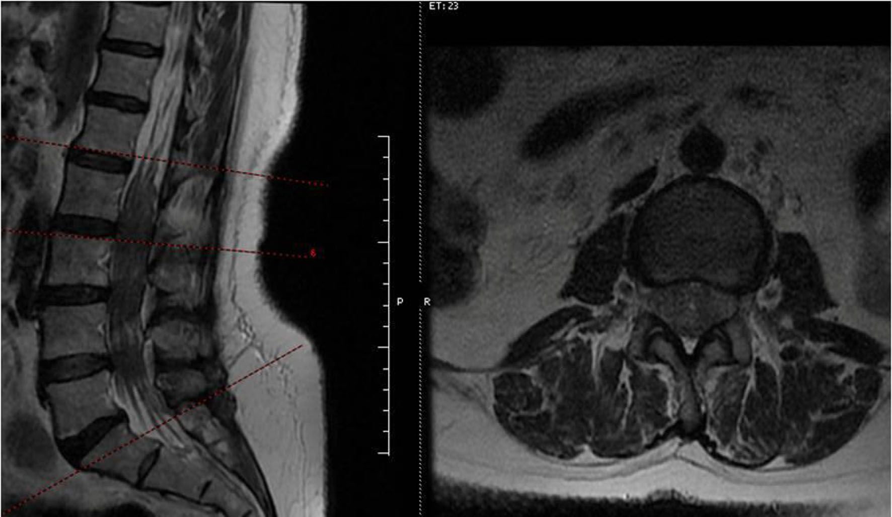 Click for large image | Figure 4. Follow-up lumbar MRI at 6 months demonstrating relatively relaxation of nerve rootlets with persistent edema. MRI: magnetic resonance imaging. |
| Discussion | ▴Top |
Arachnoiditis is an uncommon inflammatory condition of the arachnoid membrane that may result in progressive neurological deficits and significant morbidity. The etiology of arachnoiditis is heterogeneous and includes trauma, infection, hemorrhage, neoplasms, and iatrogenic factors such as spinal surgery or intrathecal interventions [1–6]. Andriuskeviciute et al recently reported a case of adhesive arachnoiditis, subarachnoid hemorrhage, and an intradural extramedullary thoracic cavernoma [1]. However, idiopathic cases without an identifiable precipitating factor have also been reported in the literature [7, 8]. In the present case, no history of trauma, surgery, infection, or intrathecal procedure was identified, supporting the diagnosis of idiopathic arachnoiditis.
MRI is the cornerstone of arachnoiditis diagnosis. Characteristic MRI findings include clumping of nerve roots, the “pseudocord” sign, obliteration of the subarachnoid space, and tethering of the spinal cord to the dorsal or ventral dura [1, 3, 9]. Arachnoiditis is classified into three groups based on MRI findings. In type I arachnoiditis, the nerve roots appear clumped and distorted. Type II is characterized by adhesion of the nerve roots to the thecal sac, resulting in the characteristic “empty thecal sac” sign. In type III arachnoiditis, both the nerve roots and the thecal sac are conglomerated into a single central soft-tissue mass within the spinal canal [10]. In the present case, lumbar MRI demonstrated a diffuse hypointense appearance occupying the spinal canal on T2-weighted images without contrast enhancement, raising suspicion for an intradural pathological process. However, definitive diagnosis could not be established radiologically, necessitating surgical exploration.
The clinical presentation of arachnoiditis is highly variable and often nonspecific, which contributes to diagnostic delay. Common symptoms include radicular pain, sensory disturbances, motor weakness, and autonomic dysfunction. In advanced cases, patients may present with severe motor deficits and gait impairment [2, 4, 5]. Our patient presented with acute-onset bilateral lower extremity weakness and sensory loss without sphincter involvement, which is relatively uncommon and may mimic other intradural or intramedullary pathologies.
Surgical management of arachnoiditis remains controversial, as outcomes are variable and the risk of symptom recurrence is high. Nevertheless, surgery may be indicated for diagnostic clarification, decompression, and lysis of adhesions in patients with progressive neurological deficits [1, 5, 7]. In our patient, surgical exploration excluded other intradural pathologies and revealed extensive nerve root edema and arachnoid adhesions, confirming the diagnosis. Adhesiolysis and duraplasty were performed to restore cerebrospinal fluid flow and reduce neural compression.
Postoperatively, the patient demonstrated significant neurological improvement, with marked recovery of motor function and reduction of sensory symptoms. Although follow-up MRI showed persistent nerve root edema, clinical improvement was sustained, suggesting that radiological findings may not always correlate with clinical outcomes. Similar observations have been reported in previous studies, emphasizing the importance of clinical evaluation during follow-up [4, 8, 10, 11].
This case highlights several important aspects of arachnoiditis. First, idiopathic arachnoiditis should be considered in the differential diagnosis of acute or subacute bilateral lower extremity weakness, especially in the absence of known risk factors. Second, MRI findings may be inconclusive, and surgical exploration may be required for definitive diagnosis. Finally, carefully selected patients may benefit from surgical intervention, with favorable clinical outcomes.
An important consideration in the present case is the potential contribution of rheumatoid arthritis (RA) to the development of arachnoiditis. RA is a chronic systemic autoimmune disease that has been associated with rare central nervous system manifestations, most notably rheumatoid meningitis and pachymeningitis. These inflammatory conditions may involve the leptomeninges and, in rare instances, extend to the spinal arachnoid space, potentially leading to fibrotic and adhesive changes.
However, in our patient, there was no clinical, laboratory, or radiological evidence of active systemic RA exacerbation or central nervous system involvement at the time of presentation. In addition, no other autoimmune or systemic inflammatory signs were identified. Therefore, although an autoimmune contribution cannot be completely excluded, a definitive causal relationship between RA and arachnoiditis could not be established in this case.
For these reasons, and in the absence of other identifiable etiological factors, the condition was considered idiopathic. Nevertheless, this case highlights the importance of considering autoimmune diseases in the differential diagnosis of arachnoiditis, even when classical risk factors are not present.
Acknowledgments
We are grateful to the patient for her cooperation and consent to share the clinical details for this report.
Financial Disclosure
No funding was received for this study.
Conflict of Interest
The authors individually declared no competing interests.
Informed Consent
Written informed consent was obtained from the patient for publication of this case report.
Author Contributions
Dr. Hakan Ak: conceptualization, primary manuscript drafting manuscript review, and editing. Dr. Fatih Durna and Dr. Sedanur Ozgul: primary manuscript drafting, data collection, and data analysis. Dr. Emine AVGIN: data collection and data analysis.
Data Availability
The authors declare that data supporting the findings of this study are available from the corresponding author upon reasonable request.
Declaration of AI Use
ChatGPT was used to assist with English language translation.
| References | ▴Top |
- Andriuskeviciute A, Mondragon-Soto MG, Penet N, Barges-Coll J. Adhesive arachnoiditis, subarachnoid hemorrhage, and intradural extramedullary thoracic cavernoma: illustrative case. J Neurosurg Case Lessons. 2024;7(13).
doi pubmed - Todeschi J, Chibbaro S, Gubian A, Pop R, Proust F, Cebula H. Spinal adhesive arachnoiditis following the rupture of an Adamkiewicz aneurysm: Literature review and a case illustration. Neurochirurgie. 2018;64(3):177-182.
doi pubmed - Safi S, Thabat A, Arshad M, Hanoun M. Arachnoiditis: a challenge in diagnosis and success in outcome: case report. Interdiscip Neurosurg. 2021;25:101219.
- Jurga S, Szymanska-Adamcewicz O, Wierzcholowski W, Pilchowska-Ujma E, Urbaniak L. Spinal adhesive arachnoiditis: three case reports and review of literature. Acta Neurol Belg. 2021;121(1):47-53.
doi pubmed - Esses SI, Morley TP. Spinal arachnoiditis. Can J Neurol Sci. 1983;10(1):2-10.
doi pubmed - Ottaiano A, De Andrade Lourencao Freddi T. Sticky spine: a review of the imaging findings for spinal arachnoiditis. Neurographics. 2022;12(3):169-175
doi - Ramesh V. Idiopathic chronic adhesive arachnoiditis presenting with paraparesis. SBV J Basic Clin Appl Health Sci. 2024;7(2):96-99.
doi - Assey EV, Sadiq AM, Swai MJ, Sadiq AM, Dekker MCJ. Idiopathic Arachnoiditis of the Cauda Equina: A Case Report from Tanzania. Case Rep Neurol. 2021;13(2):535-540.
doi pubmed - Jorgensen J, Hansen PH, Steenskov V. Chronic adhesive spinal arachnoiditis: clinical, radiological, and pathological findings. J Neurosurg. 1978;48(5):823-828.
- Kara T, Davulcu O, Ates F, Arslan FZ, Sara HI, Akin A. What happened to cauda equina fibers? Adhesive arachnoiditis. Anaesth Pain Intensive Care. 2020;24(5):555-557.
- Parenti V, Huda F, Richardson PK, Brown D, Aulakh M, Taheri MR. Lumbar arachnoiditis: Does imaging associate with clinical features? Clin Neurol Neurosurg. 2020;192:105717.
doi pubmed
This article is distributed under the terms of the Creative Commons Attribution 4.0 International License (CC BY 4.0), which permits unrestricted use, distribution, and reproduction in any medium, including commercial use, provided the original work is properly cited.
Journal of Current Surgery is published by Elmer Press Inc.