| Journal of Current Surgery, ISSN 1927-1298 print, 1927-1301 online, Open Access |
| Article copyright, the authors; Journal compilation copyright, J Curr Surg and Elmer Press Inc |
| Journal website https://jcs.elmerpub.com |
Review
Volume 16, Number 2, September 2026, pages 27-33

Contribution to Diagnostic Accuracy and Prognostic Significance of MicroRNAs in Thyroid Cancer
Kalliopi E. Stavratia  , Efstathios T. Pavlidisb , Alexandra G. Marneric , Christina Mouratidouc , Athanasios Kofinasd , Theodoros E. Pavlidisb, e
aSurgical Department, Eugenideio Hospital, Athens 11528, Greece
bSchool of Medicine, Aristotle University of Thessaloniki, 2nd Department of Propedeutic Surgery, Hippokration General Hospital, Thessaloniki 54642, Greece
cIntensive Care Unit, Hippokration General Hospital, Thessaloniki 54642, Greece
dSchool of Medicine, Aristotle University of Thessaloniki, Department of Transplantation Surgery, Hippokration General Hospital, Thessaloniki 54642, Greece
eCorresponding Author: Theodoros E. Pavlidis, School of Medicine, Aristotle University of Thessaloniki, 2nd Department of Propedeutic Surgery, Hippokration General Hospital, Thessaloniki 54642, Greece
Manuscript submitted June 5, 2026, accepted August 7, 2026, published online August 29, 2026
Short title: MicroRNAs in Thyroid Cancer
doi: https://doi.org/10.14740/jcs1049
| Abstract | ▴Top |
The incidence of thyroid cancer, the most common endocrine malignancy, has increased significantly, underscoring the need for improved diagnostic and prognostic methods. MicroRNAs (miRNAs) are small non-coding RNA molecules that regulate gene expression and play vital roles in cancer development. In thyroid cancer, they have gained attention as potential diagnostic and prognostic biomarkers. Current diagnostic methods, such as fine-needle aspiration cytology (FNAC), have limitations, particularly for indeterminate nodules (Bethesda III/IV), whereas conventional follow-up markers such as thyroglobulin (Tg) can be unreliable in certain patients. In this context, miRNAs offer a promising complementary approach. Several miRNAs, including miR-146b, miR-221, and miR-222, are consistently overexpressed in thyroid cancer and are associated with tumor presence and aggressive disease features. Circulating and exosomal miRNAs have shown good diagnostic accuracy in distinguishing malignant from benign thyroid nodules. Importantly, using multiple miRNA panels instead of single markers improves diagnostic performance and can reduce unnecessary surgeries in patients with indeterminate cytology. In addition to their diagnostic utility, miRNAs provide critical prognostic information. For instance, increased expression of miR-146b, miR-221, and miR-222 is associated with higher risk of recurrence, lymph node metastasis, and advanced tumor stages. In contrast, reduced expression of miRNAs such as miR-139-5p has been linked to persistent or progressive disease and unfavorable molecular features, including telomerase reverse transcriptase (TERT) promoter mutations. Furthermore, longitudinal changes in circulating miRNA levels may allow for early detection of disease progression, even when traditional biomarkers are inconclusive. At the molecular level, miRNAs operate within complex regulatory networks, including interactions with long non-coding RNAs and key signaling pathways to influence tumor growth and invasion. Their expression is also associated with common oncogenic mutations, such as those in the BRAF gene and RAS gene, suggesting a role in tumor heterogeneity and behavior. Recent studies highlight the potential of integrating miRNA profiles with other molecular data through multi-omics approaches, to improve risk stratification and support personalized treatment strategies. However, several challenges remain, including variability between studies, lack of standardized protocols, and limited large-scale prospective validation. In conclusion, miRNAs represent a promising and evolving class of biomarkers in thyroid cancer with direct applications in diagnosis, prognosis, and disease monitoring.
Keywords: Papillary thyroid cancer; Thyroid neoplasms; MicroRNAs; Thyroid cancer genomics; Targeted therapy
| Introduction | ▴Top |
As thyroid cancer is the most prevalent endocrine malignancy, its incidence has increased markedly—a trend potentially attributable to factors such as exposure to ionizing radiation, environmental factors, nutritional influences, and lifestyle-related stressors. It is true that this increase has been noted in many countries such as the United States, Europe, China, Australia, and South Korea; however, the mortality remained stable in most countries worldwide. A better diagnosis of micropapillary thyroid cancer (MPTC, < 1 cm) by improved new imaging methods, mainly ultrasound (US) and fine-needle aspiration cytology (FNAC), has contributed to the increasing incidence. Most experts agree that overdiagnosis can be avoided for this reason. Consequently, there is a critical need for enhanced diagnostic and prognostic methodologies [1–5]. MicroRNAs (miRNAs), which are small non-coding RNA molecules involved in regulating gene expression, are well-recognized for their pivotal role in oncogenesis. In the context of thyroid cancer, miRNAs have garnered considerable interest as promising biomarkers for both diagnosis and prognosis [3, 4]. Notably, the diagnostic sensitivity of miR-146b and miR-222 is high, validating their utility in identifying papillary thyroid cancer (PTC) [3]. Furthermore, circulating miRNAs correlate with PTC-specific clinicopathological parameters, including tumor location, tumor size, extrathyroidal extension, lymph node metastasis, the presence of the BRAF V600E gene mutation, advanced tumor node metastasis (TNM) stage, and tumor recurrence [4].
The types of thyroid cancer according to the latest World Health Organization (WHO) classification [6] are shown in Table 1.
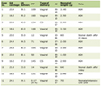 Click to view | Table 1. Types of Thyroid Cancer, Frequency, and Implicated Genes |
PTC represents the most frequently diagnosed subtype of thyroid cancer. Although the overall prognosis for most patients is favorable, the presence of lymph node metastasis is recognized as a significant high-risk factor for the development of distant metastases, which constitute the primary predictor of mortality [7]. This malignancy has the potential to evolve into more dedifferentiated and aggressive forms, including poorly differentiated papillary thyroid carcinoma (PDPTC) and anaplastic thyroid carcinoma (ATC), both of which are associated with a poor prognosis [8].
The role of miRNAs in carcinogenesis has been increasingly elucidated. These molecules have emerged as promising markers for the diagnosis and prognosis of thyroid cancer. Furthermore, integrating miRNA expression profiles with mutational status may enhance the accuracy of cytological diagnosis [9].
The genes implicated in thyroid carcinoma include the BRAF V600E gene [4, 9–12], RET gene [13–16], RAS gene [17], PTEN gene [18, 19], TP53 gene [20], PIK3CA gene [20], NTRK gene [17], and TERT gene [21], as shown in Table 1.
The diagnostic strategy for thyroid nodules includes high-frequency US, FNAC, thyroid function tests, computed tomography, and radionuclide imaging [1].
The proposed therapeutic approach for differentiated thyroid carcinoma is based on surgery, determined by tumor diameter (≥ 4 cm), combined with postoperative management. In addition to thyroidectomy, the surgical procedure often includes ipsilateral neck lymph node dissection. This may be prophylactic—performed for tumors larger than 4 cm without evidence of lymph node infiltration targeting the central compartment, or therapeutic regardless of tumor size—encompassing both central and lateral compartments in cases of advanced papillary carcinoma with cervical lymph node involvement (LNI), as well as medullary carcinoma [17, 22]. Adjuvant radioactive iodine therapy (131I) may be indicated for well-differentiated thyroid carcinoma based on histopathological findings. Additionally, thyroid-stimulating hormone (TSH) suppression therapy and thyroglobulin (Tg) measurement as a tumor marker are essential for long-term follow-up [1].
This narrative review highlights recent developments, emphasizing the emerging diagnostic and prognostic roles of miRNAs, as well as the genetic background of thyroid cancer.
| Diagnosis | ▴Top |
Current diagnostic methods for thyroid nodules, including US and FNAC, have major limitations, particularly for indeterminate cytology (Bethesda III/IV). Approximately 20–30% of thyroid nodules fall into this category, and most of these lesions are found to be benign after surgery, leading to unnecessary procedures. In addition, conventional follow-up biomarkers such as Tg can be unreliable in certain clinical settings. Calcitonin, while a well-established marker for medullary thyroid carcinoma, is not applicable to the more common differentiated thyroid cancers [1–5]. The Bethesda classification system is used to categorize thyroid cytology findings and guide clinical management decisions (Table 2).
Click to view | Table 2. Bethesda Classification of Fine-Needle Aspiration Cytology |
Notably, the greatest diagnostic uncertainty occurs in Bethesda categories III and IV, where additional molecular tools are most needed. In this context, miRNAs have emerged as promising complementary biomarkers that can improve diagnostic accuracy and reduce unnecessary surgeries [3, 4].
Circulating miRNAs have demonstrated high diagnostic performance in distinguishing malignant from benign thyroid nodules. A systematic review and meta-analysis of 35 studies reported a pooled sensitivity and specificity of 81%, with an area under the curve (AUC) of 0.88. Importantly, multi-miRNA panels significantly outperformed individual miRNAs, achieving a sensitivity of 88%, a specificity of 89%, and an AUC of 0.94 [23]. These findings suggest that combined miRNA signatures provide a more robust diagnostic approach than single biomarkers.
Among the individual miRNAs, miR-146b-5p is considered one of the most reliable diagnostic markers. Tissue-based studies have shown high diagnostic accuracy (AUC 0.94, sensitivity 89%, and specificity 87%), while circulating miR-146a-5p has also demonstrated strong performance in differentiating PTC patients from healthy controls [24, 25]. Similarly, serum miR-221-3p shows a high diagnostic value (AUC of approximately 0.87), and the diagnostic accuracy of US and FNAC can be improved when miR-222 and miR-146b are significantly elevated in malignant nodules and are used as adjunct markers [26]. Additional miRNAs, including miR-21 and miR-187-3p, are upregulated in tumor tissue, and combined panels incorporating these markers have demonstrated satisfactory diagnostic performance [27].
Furthermore, miR-146b demonstrates strong diagnostic utility for both follicular thyroid adenoma (FTA) and follicular thyroid carcinoma (FTC) [28], while the use of miRNAs miR-183-5p and miR-375-5p shows promise for the diagnosis of medullary thyroid cancer (MTC) [29].
Exosomal miRNAs represent a particularly promising subset due to their stability in circulation. A meta-analysis of 12 studies comprising 1,164 patients reported a pooled sensitivity of 82% and a specificity of 76%. Notably, specific exosomal miRNA panels have achieved excellent performance, with AUC values approaching 0.98 and sensitivity and specificity above 90% [30]. These findings highlight the potential of exosomal miRNAs as non-invasive and highly accurate diagnostic biomarkers.
One of the most clinically relevant applications of miRNAs is their use in evaluating indeterminate thyroid nodules (Bethesda III/IV). Several miRNA-based diagnostic platforms have demonstrated strong performance in this setting. A 19-miRNA next-generation sequencing panel achieved a sensitivity of 91%, a specificity of 100%, and an overall accuracy of 94% [31]. Similarly, the mir-THYpe classifier, which analyzes 11 miRNAs from fine-needle aspiration (FNA) smear slides, showed a sensitivity of 94.6%, a specificity of 81%, and a high negative predictive value (NPV) of 95.9%, comparable to established commercial platforms such as Afirma GSC and ThyroSeq v3 [32].
Other approaches, such as the ThyGeNEXT/ThyraMIR platform, combine mutation analysis with miRNA profiling and have shown improved diagnostic performance compared with mutation testing alone [33]. In addition, newer methods based on pairwise miRNA expression ratios have further improved diagnostic accuracy and reduced the number of indeterminate results, highlighting the continuous evolution in this field [34].
Compared with widely used molecular diagnostic platforms, miRNA-based panels show comparable performance. A meta-analysis of 40 studies revealed that the sensitivity and specificity of miRNA panels were similar to those of Afirma GSC and ThyroSeq v3, with the added advantages of lower costs and potentially wider accessibility [34]. These findings suggest that miRNAs can play an important role either as standalone tools or within integrated diagnostic algorithms.
At the molecular level, miRNA expression is closely linked to key oncogenic mutations. The overexpression of miR-146b, miR-221, and miR-222 is associated with BRAF V600E gene and RAS gene mutations, suggesting that combining miRNA profiles with genetic data can further improve diagnostic accuracy and better reflect tumor biology [24].
From a clinical perspective, the primary applications of miRNAs include improving the diagnosis of indeterminate nodules, enhancing the accuracy of US and cytology, and reducing unnecessary surgeries [34]. Current clinical guidelines, such as the National Comprehensive Cancer Network (NCCN) Thyroid Carcinoma Guidelines, already recommend molecular testing in selected cases of indeterminate cytology, supporting a more personalized approach to patient management [35].
Overall, miRNAs represent a promising and rapidly evolving class of diagnostic biomarkers in thyroid cancer. Their use, particularly in combination panels and in challenging clinical scenarios such as indeterminate nodules, has the potential to significantly improve diagnostic accuracy and patient management. Despite these promising results, several challenges remain, including differences in laboratory methods, and a lack of standardized cut-off values. Addressing these issues will be essential before miRNA-based diagnostics can be fully integrated into the established protocol.
| Prognosis | ▴Top |
Accumulating evidence suggests that dysregulated miRNA expression is closely associated with tumor aggressiveness, recurrence, metastasis, and survival outcomes. Among the most consistently studied miRNAs, miR-146b, miR-221, and miR-222 are upregulated in PTC and are strongly associated with adverse clinical features and poor prognosis [4, 7].
A systematic review and meta-analysis demonstrated that increased miRNA expression is significantly associated with worse overall survival (hazard ratio (HR) = 5.94, 95% confidence interval (CI): 2.73–12.90) and disease- or recurrence-free survival (HR = 1.58, 95% CI: 1.08–2.32) [33]. Similarly, in another meta-analysis, miR-146b, miR-222, and miR-221 were identified as the strongest individual predictors of recurrence, with odds ratios (ORs) of 9.11, 6.56, and 3.88, respectively [36]. These findings support the role of specific miRNAs as reliable markers for risk stratification in patients with thyroid cancer.
In addition to survival outcomes, several miRNAs can predict key clinicopathological features. For example, miR-146b-5p and miR-221-3p have demonstrated a strong predictive value for lymph node metastasis and disease recurrence. A CRISPR-based blood assay revealed that these miRNAs performed well in predicting LNI (AUCs of 0.816 and 0.740, respectively) and recurrence (AUCs of 0.921 and 0.756), outperforming traditional systems such as TNM staging and American Thyroid Association (ATA) risk classification [37]. Similarly, miR-222-3p has been identified as an independent predictor of extrathyroidal extension and advanced tumor stage, further supporting its value in identifying high-risk disease [38].
In addition to their diagnostic role, miRNAs provide important prognostic information in thyroid cancer. Increased expression of miR-146b, miR-221, and miR-222 has been consistently associated with increased risks of recurrence, lymph node metastasis, and more advanced tumor stage [36]. In contrast, reduced expression of specific miRNAs, such as miR-139-5p, has been linked to persistent or progressive disease, as well as unfavorable molecular features, including telomerase reverse transcriptase (TERT) promoter mutations [21].
Furthermore, dynamic changes in circulating miRNA levels may allow for early detection of disease progression, even when conventional biomarkers such as Tg are inconclusive [25]. This highlights the potential role of miRNAs not only as prognostic markers, but also as tools for dynamic disease surveillance.
Additional miRNAs have also been associated with disease progression. High expression of miR-136, miR-21, and miR-127 has been linked to distant metastasis and recurrent or persistent disease, suggesting their involvement in tumor dissemination [36]. The expression of miR-155 is also upregulated in thyroid malignancies, with a weak association with LNI, although further validation is required [39].
At the molecular level, miRNAs are involved in complex regulatory networks, including interactions with long non-coding RNAs and key signaling pathways that influence tumor growth and invasion. Their expression is also associated with common oncogenic mutations, such as those in the BRAF gene and RAS gene, suggesting that they play a role in tumor heterogeneity and tumor behavior [24]. These findings indicate that miRNAs can act together with genetic alterations to drive tumor progression and could improve prognostic accuracy when used in combination.
Recent studies have also highlighted the potential of integrating miRNA profiles with other molecular data through multi-omics approaches, to improve risk stratification and support more personalized treatment strategies. This integrated approach can better capture the biological complexity of thyroid cancer and allow for a more accurate prediction of disease outcomes [40].
Epigenetic markers—including promoter hypermethylation of tumor suppressor genes, global DNA hypomethylation, and specific miRNAs such as miR-146b, miR-221, and miR-375—have emerged as promising indicators of aggressive disease progression in well-differentiated thyroid cancer [41].
LNI is a critical prognostic factor associated with unfavorable survival outcomes in MTC patients. The presence of desmoplastic stroma and elevated expression levels of miR-21 have been identified as robust predictors of LNI, potentially guiding surgical decision-making regarding the extent of cervical lymph node dissection in patients with large, sporadic MTCs lacking preoperative evidence of LNI [42].
Additionally, the expression of miR-224 is upregulated in RAS-mutated MTCs and is associated with an improved patient prognosis, suggesting its potential role as an independent prognostic biomarker in this disease [13].
Furthermore, miR-21 modulates the expression of programmed cell death 4 (PDCD4), with the miR-21/PDCD4 signaling axis demonstrating significant correlations with clinicopathological features and patient prognosis [14].
Importantly, the prognostic value of miRNAs can have implications for targeted therapy. Specific miRNA expression profiles are associated with key oncogenic pathways and molecular subtypes, which can influence treatment response. For example, the interaction of miRNAs with signaling pathways related to BRAF gene and RAS gene mutations suggests that they could help identify patients who will benefit from targeted therapies. However, the role of miRNAs in guiding therapeutic decisions is still under investigation and requires further clinical validation [40, 41, 43].
MicroRNA might be another tool to select patients more appropriately. This method is promising, but more documentation is needed. The studies show quite contradictory results especially concerning the cut-off values and the standardization of the protocols, as mentioned above [44, 45].
Overall, miRNAs represent a promising class of prognostic biomarkers in thyroid cancer. They provide important information regarding tumor behavior, risk of recurrence, and likelihood of disease progression, often complementing or even exceeding traditional clinical systems. Despite these encouraging findings, several limitations remain, including inter-study variability, a lack of standardized methodologies, and limited large-scale prospective validation. Further research is needed to establish standardized protocols and confirm the clinical utility of miRNAs in routine clinical practice.
| Conclusions | ▴Top |
miRNAs represent a rapidly evolving, promising class of biomarkers in thyroid cancer, with potential applications in diagnosis, prognosis, and disease monitoring. The combined analysis of miRNA expression profiles and mutational status may enhance the accuracy of cytological diagnosis. These molecules have emerged as valuable predictors of tumor progression and aggressiveness. Furthermore, miRNAs provide a more precise assessment of the risk of adverse clinical outcomes and may inform the selection of targeted therapeutic strategies. Incorporating miRNAs into clinical protocols has the potential to improve patient management, particularly in complex diagnostic and follow-up contexts. However, several challenges remain, including variability across studies, a lack of standardized analytical methods, and a limited number of large-scale prospective studies assessing clinical outcomes.
Acknowledgments
None to declare.
Financial Disclosure
No funding was received in regard to the production of this paper.
Conflict of Interest
The authors declared no potential conflict of interest.
Author Contributions
KES, ETP, and AM designed the research and analyzed the data. AK and CM performed the research, contributed new analytic tools, evaluated the data, and reviewed the paper. TEP analyzed the data review and approved the paper. All authors have read and approved the final manuscript.
Data Availability
All data generated or analyzed during this study are included in this published article, and further inquiries should be directed to the corresponding author.
AI Use Declaration
The manuscript was not AI-generated. The content was written and developed by the authors. AI tools were used solely for language editing and polishing to improve clarity and readability of the manuscript. The manuscript underwent standard academic revisions by the authors.
Abbreviations
ATA: American Thyroid Association; ATC: anaplastic thyroid carcinoma; AUC: area under the curve; AUS/FLUS: atypia undetermined significance/follicular lesion undetermined significance; Bethesda III/IV: indeterminate thyroid nodules; CI: confidence interval; FNA: fine-needle aspiration; FNAC: fine-needle aspiration cytology; FTA: follicular thyroid adenoma; FTC: follicular thyroid carcinoma; FN/SFN: follicular neoplasm/suspicious follicular neoplasm; HR: hazard ratio; 131I: adjuvant radioactive iodine therapy; LNI: lymph node involvement; miRNAs: microRNAs; MTC: medullary thyroid cancer; MPTC: micropapillary thyroid cancer; NCCN: National Comprehensive Cancer Network; NPV: negative predictive value; OR: odds ratio; PTC: papillary thyroid cancer; PDCD4: programmed cell death 4; PDPTC: papillary thyroid carcinoma; Tg: thyroglobulin; TERT: telomerase reverse transcriptase; TNM: tumor node metastasis; TSH: thyroid-stimulating hormone; US: ultrasound; WHO: World Health Organization
| References | ▴Top |
- Wang Y, Liu J, Zhang CX, Gu GL. Thyroid cancer in Chinese military flight personnel: Characteristics and management advances. World J Clin Oncol. 2026;17(2):115364.
doi pubmed - Hsu CW, Hsueh C, Lu YL, Hsu CJ, Wong RJ, Lin SF. Risk factors and outcomes of metastatic poorly differentiated thyroid carcinoma. World J Clin Cases. 2025;13(25):105204.
doi pubmed - Dean B, Geropoulos G, Richardson-Jones T, Fornasiero M, Papapanou M, Konstantinidis C, Madouros N, et al. Diagnostic accuracy of circulating miR-146, miR-221 and miR-222 in papillary thyroid cancer: A systematic review and meta-analysis. World J Clin Cases. 2025;13(27):104916.
doi pubmed - Geropoulos G, Psarras K, Papaioannou M, Giannis D, Meitanidou M, Kapriniotis K, Symeonidis N, et al. Circulating microRNAs and clinicopathological findings of papillary thyroid cancer: a systematic review. In Vivo. 2022;36(4):1551-1569.
doi pubmed - Tziona EE, Emmanouilidou A, Stouras I, Omar CT, Koloka M, Savva A, Dimitrakopoulou K, et al. Thyroid carcinoma of the pyramidal lobe: A narrative review of reported cases. World J Clin Cases. 2026;14.
doi - Jung CK, Bychkov A, Kakudo K. Update from the 2022 World Health Organization classification of thyroid tumors: a standardized diagnostic approach. Endocrinol Metab (Seoul). 2022;37(5):703-718.
doi pubmed - Tekin E, Oflas D, Badak B, Kebapci N, Akalin A, Dundar E. Prognostic significance of Lin28A protein expression in papillary thyroid carcinoma. Indian J Pathol Microbiol. 2025;68(3):486-491.
doi pubmed - Dadafarin S, Carnazza M, Islam HK, Moscatello A, Tiwari RK, Geliebter J. Noncoding RNAs in papillary thyroid cancer: interaction with cancer-associated fibroblasts (CAFs) in the tumor microenvironment (TME) and regulators of differentiation and lymph node metastasis. Adv Exp Med Biol. 2021;1350:145-155.
doi pubmed - Denaro M, Ugolini C, Poma AM, Borrelli N, Materazzi G, Piaggi P, Chiarugi M, et al. Differences in miRNA expression profiles between wild-type and mutated NIFTPs. Endocr Relat Cancer. 2017;24(10):543-553.
doi pubmed - Pamedytyte D, Simanaviciene V, Dauksiene D, Leipute E, Zvirbliene A, Sarauskas V, Dauksa A, et al. Association of MicroRNA expression and BRAF(V600E) mutation with recurrence of thyroid cancer. Biomolecules. 2020;10(4):625.
doi pubmed - Han PA, Kim HS, Cho S, Fazeli R, Najafian A, Khawaja H, McAlexander M, et al. Association of BRAF V600E mutation and MicroRNA expression with central lymph node metastases in papillary thyroid cancer: a prospective study from four endocrine surgery centers. Thyroid. 2016;26(4):532-542.
doi pubmed - Huang Y, Liao D, Pan L, Ye R, Li X, Wang S, Ye C, et al. Expressions of miRNAs in papillary thyroid carcinoma and their associations with the BRAFV600E mutation. Eur J Endocrinol. 2013;168(5):675-681.
doi pubmed - Cavedon E, Barollo S, Bertazza L, Pennelli G, Galuppini F, Watutantrige-Fernando S, Censi S, et al. Prognostic impact of miR-224 and RAS mutations in medullary thyroid carcinoma. Int J Endocrinol. 2017;2017:4915736.
doi pubmed - Pennelli G, Galuppini F, Barollo S, Cavedon E, Bertazza L, Fassan M, Guzzardo V, et al. The PDCD4/miR-21 pathway in medullary thyroid carcinoma. Hum Pathol. 2015;46(1):50-57.
doi pubmed - Mian C, Pennelli G, Fassan M, Balistreri M, Barollo S, Cavedon E, Galuppini F, et al. MicroRNA profiles in familial and sporadic medullary thyroid carcinoma: preliminary relationships with RET status and outcome. Thyroid. 2012;22(9):890-896.
doi pubmed - Yip L, Kelly L, Shuai Y, Armstrong MJ, Nikiforov YE, Carty SE, Nikiforova MN. MicroRNA signature distinguishes the degree of aggressiveness of papillary thyroid carcinoma. Ann Surg Oncol. 2011;18(7):2035-2041.
doi pubmed - Pavlidis ET, Pavlidis TE. Role of prophylactic central neck lymph node dissection for papillary thyroid carcinoma in the era of de-escalation. World J Clin Oncol. 2023;14(7):247-258.
doi pubmed - Guo K, Wang J, Shu L, Zhou G. MiR-200c promotes papillary thyroid cancer cell proliferation, migration, and invasion by downregulating PTEN. Tissue Cell. 2021;73:101647.
doi pubmed - Nikiforova MN, Nikiforov YE. Molecular diagnostics and predictors in thyroid cancer. Thyroid. 2009;19(12):1351-1361.
doi pubmed - Pavlidis ET, Galanis IN, Pavlidis TE. Update on current diagnosis and management of anaplastic thyroid carcinoma. World J Clin Oncol. 2023;14(12):570-583.
doi pubmed - Martinez-Puente N, Ruz-Caracuel I, Leandro-Garcia LJ, Pian-Arias H, Vega-Corral Z, Leton R, Radu R, et al. Expression of Homo Sapiens (Hsa)-miR-139-5p as a clinically feasible prognostic marker for differentiated thyroid cancer. Lab Invest. 2025;105(10):104199.
doi pubmed - Pavlidis E, Sapalidis K, Chatzinikolaou F, Kesisoglou I. Medullary thyroid cancer: molecular factors, management and treatment. Rom J Morphol Embryol. 2020;61(3):681-686.
doi pubmed - Xu SL, Tian YY, Zhou Y, Liu LQ. Diagnostic value of circulating microRNAs in thyroid carcinoma: a systematic review and meta-analysis. Clin Endocrinol (Oxf). 2020;93(4):489-498.
doi pubmed - Matos ML, Pinto M, Alves M, Canberk S, Goncalves A, Bugalho MJ, Papoila AL, et al. Cyto-histological profile of MicroRNAs as diagnostic biomarkers in differentiated thyroid carcinomas. Genes (Basel). 2024;15(3):389.
doi pubmed - Verrienti A, Pecce V, Grani G, Del Gatto V, Barp S, Maranghi M, Giacomelli L, et al. Serum microRNA-146a-5p and microRNA-221-3p as potential clinical biomarkers for papillary thyroid carcinoma. J Endocrinol Invest. 2025;48(3):619-631.
doi pubmed - Samir Assaad R, Saad Ragab W, El Tayfi R, El Sheredy AG. Circulating microRNA-222 and microRNA-146b: a potential differentiating tool between benign and malignant thyroid nodules. Scand J Clin Lab Invest. 2025;85(7):581-589.
doi pubmed - Baig RM, Afzal A, Khan R, Kainat N. Evaluation of expressional deregulations of micro RNAs and their potential role as molecular biomarkers in the diagnosis and prognosis of papillary thyroid carcinoma. J Clin Oncol. 2025;43(16_suppl):e18147.
doi - Sheikholeslami S, Shabani N, Shivaee S, Tavangar SM, Yeganeh M, Hedayati M, Lotfi J, et al. Overexpression of mir-129-1, miR-146b, mir-183, and mir-197 in follicular thyroid carcinoma and adenoma tissues. Mol Cell Probes. 2020;51:101536.
doi pubmed - Febrero B, Ros-Madrid I, Revilla-Nuin B, Abellan M, Ruiz-Manzanera JJ, Gomez J, Rodriguez JM. Could microRNA analysis help in the management of medullary thyroid cancer? Cancers (Basel). 2025;17(4):629.
doi pubmed - Toraih EA, Elshazli RM, Trinh LN, Hussein MH, Attia AA, Ruiz EML, Zerfaoui M, et al. Diagnostic and prognostic performance of liquid biopsy-derived exosomal MicroRNAs in thyroid cancer patients: a systematic review and meta-analysis. Cancers (Basel). 2021;13(17):4295.
doi pubmed - Mazeh H, Deutch T, Karas A, Bogardus KA, Mizrahi I, Gur-Wahnon D, Ben-Dov IZ. Next-generation sequencing identifies a highly accurate miRNA panel that distinguishes well-differentiated thyroid cancer from benign thyroid nodules. Cancer Epidemiol Biomarkers Prev. 2018;27(8):858-863.
doi pubmed - Labourier E, Shifrin A, Busseniers AE, Lupo MA, Manganelli ML, Andruss B, Wylie D, et al. Molecular testing for miRNA, mRNA, and DNA on fine-needle aspiration improves the preoperative diagnosis of thyroid nodules with indeterminate cytology. J Clin Endocrinol Metab. 2015;100(7):2743-2750.
doi pubmed - Finkelstein SD, Sistrunk JW, Malchoff C, Thompson DV, Kumar G, Timmaraju VA, Repko B, et al. A retrospective evaluation of the diagnostic performance of an interdependent pairwise MicroRNA expression analysis with a mutation panel in indeterminate thyroid nodules. Thyroid. 2022;32(11):1362-1371.
doi pubmed - Silaghi CA, Lozovanu V, Georgescu CE, Georgescu RD, Susman S, Nasui BA, Dobrean A, et al. Thyroseq v3, Afirma GSC, and microRNA panels versus previous molecular tests in the preoperative diagnosis of indeterminate thyroid nodules: a systematic review and meta-analysis. Front Endocrinol (Lausanne). 2021;12:649522.
doi pubmed - Haddad RI, Bischoff L, Applewhite M, Bernet V, Blomain E, Brito M, Busaidy NL, et al. NCCN Guidelines(R) insights: thyroid carcinoma, Version 1.2025. J Natl Compr Canc Netw. 2025;23(7):e250033.
doi pubmed - Silaghi CA, Lozovanu V, Silaghi H, Georgescu RD, Pop C, Dobrean A, Georgescu CE. The prognostic value of MicroRNAs in thyroid cancers-a systematic review and meta-analysis. Cancers (Basel). 2020;12(9):2608.
doi pubmed - Toraih EA, Ruiz E, Ning B, Tortelote GG, Hilliard S, Moroz K, Hu T, et al. Chromatin-accessible miRNA regulons driving thyroid tumorigenesis and progression. J Am Coll Surg. 2023;236(4):732-750.
doi pubmed - Stojanovic S, Dobrijevic Z, Selemetjev S, Doric I, Jankovic Miljus J, Zivaljevic V, Isic Dencic T. MiR-203a-3p, miR-204-3p, miR-222-3p as useful diagnostic and prognostic tool for thyroid neoplasia spectrum. Endocrine. 2023;79(1):98-112.
doi pubmed - Ibrahim DR, El-Kholany SH, Ghattas MH, El-Nahas M, Abd El-Fadeal NM. Circulating MiRNAs in thyroid cancer: prognostic promise of miR-155 and limitations of miR-429. Mol Biol Rep. 2025;53(1):147.
doi pubmed - Wang Z, Han Q, Hu X, Wang X, Sun R, Huang S, Chen W. Multi-omics clustering analysis carries out the molecular-specific subtypes of thyroid carcinoma: implicating for the precise treatment strategies. Genes Immun. 2025;26(2):137-150.
doi pubmed - Sloneva N, Kaidarova D, Kaibarov M. Prognostic stratification of highly differentiated thyroid cancer based on molecular genetic studies. Asian Pac J Cancer Prev. 2026;27(2):757-767.
doi pubmed - Aubert S, Berdelou A, Gnemmi V, Behal H, Caiazzo R, D'Herbomez M, Pigny P, et al. Large sporadic thyroid medullary carcinomas: predictive factors for lymph node involvement. Virchows Arch. 2018;472(3):461-468.
doi pubmed - Golla U, Sesham K, Dallavalasa S, Manda NK, Unnam S, Sanapala AK, Nalla S, et al. ABHD11-AS1: an emerging long non-coding RNA (lncRNA) with clinical significance in human malignancies. Noncoding RNA. 2022;8(2):21.
doi pubmed - Sonmez G, Unluturk U. A systematic review of emerging RNA markers in thyroid fine needle aspiration cytology samples: advancements and challenges. Endocrine. 2025;89(2):365-379.
doi pubmed - Ferraz C, Cunha GB, de Oliveira MMB, Tenorio LR, Cury AN, Padovani RDP, Ward LS. The diagnostic and prognostic role of miR-146b-5p in differentiated thyroid carcinomas. Front Endocrinol (Lausanne). 2024;15:1390743.
doi pubmed
This article is distributed under the terms of the Creative Commons Attribution 4.0 International License (CC BY 4.0), which permits unrestricted use, distribution, and reproduction in any medium, including commercial use, provided the original work is properly cited.
Journal of Current Surgery is published by Elmer Press Inc.